Flutemetamol Useful in Finding Neuritic Plaques
Finding brain beta amyloid is usefull in assessing Alzheimer's Disease. In a report in JAMA Neurology, Craig Curtis, MD, and colleagues report on a multicenter PET imaging study on terminally ill dementia patients using flutemetamol injections tagged with radioactive fluorine 18. In cases where autopsy was later possible, the team assessed how accurate the tests had been in diagnosing Alzheimer's.


Finding brain beta amyloid plaques is usefull in assessing Alzheimer's Disease (AD). In an article in JAMA Neurology, Craig Curtis, MD and colleagues report on a multicenter PET imaging study on terminally ill dementia patients using flutemetamol injections tagged with radioactive fluorine 18. In cases where autopsy was later possible, the team assessed how helpful the scans had been in diagnosing AD.
The primary object of the study was to determine how sensitive these images of brains of these patients were in detecting plaques. A secondary objective was to judge the specificity of the scans and see if that changed with CT images were available during the PET image review.
The study was done in the US and the UK. Based on clinical signs, atients were classified for statistical purposes as having early AD even though researchers expected not all would turn out to have it.
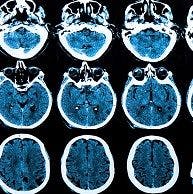
The patients were given MRI or head CT scans. Then they were injected with flutemetamol tagged with a radioactaive substance and given a PET scan.
The team recruited nuclear medicine specialists to read the scans. The images were of 5 brain regions, the combined lateral frontal cortex and anterior cingulate, combined posterior cingulate an precuneus, insula, lateral temporal lobes, and striatal regions.
In all patients the white matter of their brains showed nonspecific uptake of flutemetamol. The key to diagnosis was what part or parts of the grey matter did that--an indication of plaques. If none the grey matter showed uptake, the test was negative.
On autopsy of the patients who died, slides were made so researchers could look for microsopic signs of plaques in the various brain regions. There were 68 brains examined post mortem.
Of these 25 brains were negative for plaques and 43 were positive with plague density ranging from "none" to "frequent".
Comparing those patient-specific histopathological results to the diagnoses made before death based on the scans, the team found that when scan readers interpreted PET scans with CT images sensitivity and specificity improved but only slightly.
The scan readers made 8 wrong diagnoses: false negative interpretations of 6 of the 43 patients who had significant plaques and 2 false positive findings in the 25 patients who had no plaques.
The authors suggest that error rate was partly because one of the physicians reading the scans had not receieved enough training, a problem they said could be corrected. They also said the technique offered good image quality.
There are two other amyloid PET scan agents approved in the US and in Europe, but none have been evaluated in head-to-head trials, the researchers said.
The researchers said their study was useful in that it showed "In vivo detection of brain beta amyloid may help increase diagnostic accuracy in cognitively impaired patientss compared with clinical diagnosis alone," and that they learned that "flutemetamol injection was safe and had high sensitivity and specificity" in the group studied."