MRIs May Be Able to Detect Patients at Risk for Developing Multiple Sclerosis
Results from a study published in the International Journal of MS Care suggest that magnetic resonance imaging may be used to identify asymptomatic patients who are at higher risk for developing multiple sclerosis.

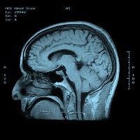

Patients with magnetic resonance images (MRIs) suggestuve of multiple sclerosis (MS) were more likely to develop MS, even if they did not present with MS symptoms at the time, according to research published in the International Journal of MS Care.
Researchers from Henry Ford Hospital in Detroit, Michigan studied 30 patients (90 percent female; 77 percent white) at an MS clinic who had abnormal MRI readings. The patients’ mean age was 43.3 years and they showed abnormal brain MRI and were observed longitudinally, every 6 or 12 months. Cerebrospinal fluid (CSF) and blood sample data was collected or assessed for evidence of MS. Each participant had also undergone neurological evaluation, but no abnormal findings were discovered. The lesions discovered in the MRIs were presented to neurologists to determine if they were suggestive of MS. At the end of the study period, participants were categorized into having MS or not having MS.
Half of the patients (50 percent) had presenting MRI scans that were considered by the researchers strongly suggestive of MS. Of those 15 participants, 7 were diagnosed with MS, and 8 were not given a MS diagnosis during the follow up period. Participants who developed MS during the follow up period had demonstrated earlier symptoms of partial myelitis (4 patients), brain stem syndrome (1 patient), and optic neuritis (2 patients). No patient whose MRI demonstrated no signs of MS developed clinical manifestations of MS.
Of the 15 patients whose CSF was analyzed, 6 had abnormal findings; 4 of those patients with abnormal findings were diagnosed with MS. One of the 9 patients with normal CSF testing developed MS during the follow up period. CSF findings of 9 patients showed 22 percent with MRI lesions suggestive of MS, while the remaining 83 percent of patients with abnormal CSF findings.
Symptoms of MS mentioned by the researchers were headache (the most common at 50 percent), dizziness (7 percent), blurry vision (7 percent), and paresthesia (7 percent). Headaches were not a significant symptom of MS diagnosis — 20 percent and 26 percent of participants with headaches were diagnosed and did not receive a MS diagnosis, respectively. Lesions were also not a suggestive symptom of MS in participants with MRIs. The researchers noted, however, the sample size was too small to test the association between MS diagnosis and presence of MRI lesions suggestive of MS.
“None of the participants without symptoms and MRI findings suggestive of MS developed MS during follow-up,” the authors concluded. “This finding may help physicians to decide whether to order follow-up brain MRI scans for individuals who lack symptoms and MRI findings suggestive of MS. Particularly in the United States, brain MRI is frequently performed because of a variety of symptoms, leading to potential overuse of this diagnostic procedure.”
The researchers highlight the importance of further, larger-scale research to determine specific characteristics that increase the likelihood of developing MS. They believe this will help counseling patients, planning imaging follow up, and identifying treatment options.