MRIs Useful in Tracking Depression in MS Patients
MRI technology can allow scientists to assess the development of depression and treatment responses over time in multiple sclerosis patients.


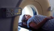
Magnetic resonance imaging (MRI) can be utilized to predict depression treatment responses in multiple sclerosis (MS) patients, according to a presentation at the 2014 Annual Meeting of the Consortium of Multiple Sclerosis Centers (CMSC) and the Sixth Cooperative Meeting with Americas Committee for Treatment and Research in Multiple Sclerosis (ACTRIMS), held May 28-31, in Dallas, Texas.
In his presentation, Anthony Feinstein, MBBCh, MPhil, PhD, associate scientist at the Sunnybrook Health Sciences Centre in Toronto, Ontario, Canada, stressed that MRI technology can allow scientists to assess the development of depression and treatment responses over time in MS patients. Lifetime prevalence of depression in patients attending MS clinics approaches 50%, which increases in a community-based sample, Feinstein told his presentation audience.
Feinstein believes MRI scans can give clues to the structural brain changes linked to depression in MS patients who suffer from pseudobulbar affect. As many as 10% of MS patients suffer from pseudobulbar affect, which is categorized by episodes of uncontrolled crying but are absent emotions like sadness, laughing, or happiness.
In his study on pseudobulbar affect, Feinstein and his research team concluded one in 10 MS patients suffer from the affect, and it occurs more frequently in severely disabled patients, generally with long-standing disease. Patients were not more depressed or anxious compared to the control group, but had a greater decline in IQ.
In a more recent study on pseudobulbar affect in MS patients tracked with MRIs, Feinstein et. al., concluded MS patients have distinct brain lesions when compared to MS controls without pseudobulbar affect. The study including 14 patients with MS and pseudobulbar affect, and 14 control patients matched on demographic, disease course, and disability variables.
Feinstein detailed what depression looks like in MS patients, and said it could be a combination of 5 or more of any of the following symptoms: feeling depressed most of the day; diminished pleasure in activities; appetite change; significant weight loss or gain; psychomotor agitation or retardation; fatigue or loss of energy nearly every day; worthlessness; excessive guilt; inappropriate guilt; having insomnia or hypersomnia nearly every day; diminished ability to think or concentrate; or recurrent thoughts of death 5 or more times during the same 2-week period.
Feinstein and others believe the hippocampus plays an important part in determining depression in MS patients. A study from Cedars-Sinai concluded a reduced size of the right hippocampus is linked to MS patients with symptoms of depressive affect, such as depressed mood and loss of interest. Another study from the University of Calgary is working to develop a new MRI technique that would track small changes in patterns of MS patients’ brain. The researchers in that study want to create an algorithm to identify texture homogeneity or changes in the tissue in MS patients’ brains.