Modern Technology Reveals Atherosclerosis Was Prevalent in Ancient Populations
Investigators in the expanded HORUS trial using CT scans of ancient mummies from multiple populations and geographic regions report finding evidence of probable or definite atherosclerosis in nearly one-third of samples.


Atherosclerosis has long been presumed to be a product of our modern diets and increasingly sedentary lifestyles, a supposition seemingly confirmed by increasing rates of atherosclerosis seen in developing countries as they westernize.
However, new evidence shows that atherosclerosis was fairly common in a wide range of ancient civilizations. A highly-anticipated featured research trial presented Sunday, March 10, ACC.13, the 62nd Annual Scientific Session & Expo of the American College of Cardiology, examined data from over 100 mummies from around the world for their atherosclerosis burden, as a follow-up to a 2011 CT study of Egyptian mummies. Published at the Lancet online, the study’s title comes from Horus, the Egyptian deity.
“Heart disease is a serial killer that has been stalking mankind for 4,000 years,” said lead investigator Randall Thompson, MD, of St. Luke’s Mid-America Heart Institute in Kansas City, MO, who presented the study.
When the previous study was published, questions arose about the probable homogeneity of the Egyptian samples, which came from a wealthy, inactive population eating rich diets. This latest study broadened the investigation, gathering more expertise and obtaining for CT scan 137 mummies of different classes, of which 76 were ancient Egyptians, 51 Peruvian, 5 ancient pueblans/Anasazi, and 5 Unangans from the Aleutian islands (who would’ve lived a traditional hunter/gatherer lifestyle, hunting from kayaks, etc).
Interpreting results by consensus, seven cardiologists undertook expeditions to Peru and Egypt, finding atherosclerosis in 34% of the mummies, in all four geographical populations: 29 (38%) Egyptians, 13 (25%) Peruvians, two (40%) Anasazi, and three (60%) Unangans. Mummies were from periods ranging from 1500 BC-1500 AD in all except the Alaskan mummies (ca. 1756-1930 AD).
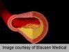
Researchers used whole-body CT scans to look for atherosclerosis, in the form of arterial wall calcification. Atherosclerosis was present in the aorta in 28 (20%) mummies, iliac or femoral arteries in 25 (18%), popliteal or tibial arteries in 25 (18%), carotid arteries in 17 (12%), and coronary arteries in six (4%). Of the five vascular beds examined, atherosclerosis was present in 1-2 beds in 34 (25%) mummies, in 3-4 beds in 11 (8%), and in all five vascular beds in two (1%). Age at death positively correlated with atherosclerosis (about 43 years for atherosclerotic mummies versus 32 years for healthy ones) and with the number of arterial beds involved (mean age of 32 years for mummies without atherosclerosis, 42 years for those with atherosclerosis in one or two beds, and 44 years for those with atherosclerosis in 3-5 beds).
These calcification findings correlate with more current autopsy study results, and do not contradict evidence that atherosclerotic disease is caused by genetic and environmental interaction associated with classic risk factors. Even with different diets, lifestyles, and cultures, disease appears inherent in human aging. Age was a predictor of the disease, found more frequently in the longer-lived, and also a predictor of increasing severity of the disease. Its presence in pre-modern humans raises the possibility of a more basic predisposition to the disease, and other factors not yet understood.
This is still a work in progress, with much more data yet to come‑‑perhaps more imaging in more populations, and tests for genetics and risk factors. The evolving area of paleocardiology, a noninvasive way to study, dates back over 100 years. The study topic has generated tremendous interest in the cardiology field, and may compel contemporary reexamination of CV risk.