Meniscus Extrusion Predicts Radiographic Osteoarthritis Change Better Than Cartilage Volume Loss
Although change in knee joint space narrowing depicted on radiographs is considered the gold standard for monitoring osteoarthritis progression and knee cartilage volume loss, the method doesn't account for extrusion of the medial meniscus, which new research suggests can better predict radiographic osteoarthritis change over time.


Although change in knee joint space narrowing depicted on radiographs is considered the gold standard for monitoring osteoarthritis progression and knee cartilage volume loss, the method doesn’t account for extrusion of the medial meniscus, which new research suggests can better predict radiographic osteoarthritis change over time.
For their poster presented at the American College of Rheumatology 2013 Annual Meeting in San Diego, CA, Louisa Chou, MBBS, of the Menzies Research Institute Tasmania at the University of Tasmania in Australia, and co-authors from Department of Epidemiology and Preventive Medicine at Monash University and the Osteoarthritis Research Unit at the University of Montreal Hospital Research Centre evaluated a cohort of 220 middle-aged patients with a higher risk of osteoarthritis at baseline, 2 years, and 10 years in an effort to “quantitate the correlation between changes in medial meniscal extrusion and knee cartilage volume loss over 2 years and radiographic osteoarthritis change over 10 years.”
According to the researchers, about 50% of the subjects were the adult offspring of patients who had knee replacement surgery performed for knee osteoarthritis, while the rest of the study population was comprised of age-, sex-, and body mass index (BMI)-matched control subjects.
At baseline, the participants’ mean medial tibia cartilage volume was 2,234 mm3 with an average annual knee cartilage volume loss of 2.5% over 2 years. Additionally, 8% of the patients had medial meniscal extrusion at baseline, while 3% had an incident meniscal extrusion over 2 years.
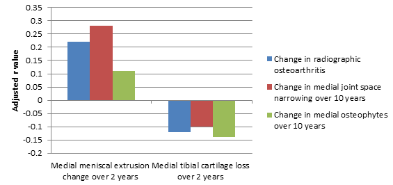
Using a Spearman’s rank-order correlation analysis to observe the association of changes in medial tibia cartilage and medial meniscal extrusion loss with changes in radiographic osteoarthritis, joint space narrowing, and osteophytes, which are more commonly known as bone spurs, the poster authors found that “an increase in medial meniscal extrusion over 2 years predicted a change in radiographic osteoarthritis over 10 years in adjusted analysis (r= 0.22, P= 0.003), an increase in medial joint space narrowing (r= 0.30, P< 0.001) and an increase in medial osteophytes (r= 0.15, P= 0.046).”
However, looking at the change in medial tibia cartilage over 2 years, the researchers noted that it “did not correlate with a change in radiographic osteoarthritis over 10 years (r= -0.12, P= 0.096) or a change in medial joint space narrowing (r= -0.10, P= 0.182).” Furthermore, medial tibia cartilage change over 2 years “correlated weakly with change in medial osteophytes (r= -0.14, P= 0.045),” they added (Figure 1).


Nevertheless, the poster authors said that their findings taken together as a whole suggest that “a change in medial meniscal extrusion — despite being quite rare — is more strongly correlated with subsequent change in radiographic osteoarthritis than cartilage volume loss.” Therefore, they concluded that “radiographic osteoarthritis is a composite measure of joint pathology and does not primarily or sufficiently reflect cartilage loss.”


