Multiple Sclerosis: Lesions on T2 Indicate Poor Prognosis
Patients with relapsing remitting multiple sclerosis (RRMS) who have T2 spinal cord (SC) lesions detected through magnetic resonance imaging (MRI) early in the course of the disease are likely to have a worse prognosis, according to recent research.

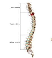

Patients with relapsing remitting multiple sclerosis (RRMS) who have T2 spinal cord (SC) lesions detected through magnetic resonance imaging (MRI) early in the course of the disease are likely to have a worse prognosis, according to recent research. A paper authored by Emanuele D’Amico of the Neurology Department at the University of Catania in Spain, and colleagues, was published in the journal Multiple Sclerosis Journal Experimental Translational Clinical describes the findings.
Describing their objective, the authors say, “In the current study, we studied a large cohort of people with RRMS (pwRRMS) to determine whether the presence of lesions in SC T2 MRI at the diagnosis of pwRRMS could predict a worse prognosis in terms of disability.” In order to investigate, the researchers reviewed collected data from multiple care centers from January, 1995 to December 2007. There were 239 participants.
“We found that the presence of SC lesions detected early at the time of diagnosis (within two years from disease onset) and EDSS [Expanded Disability Status Score] level at our baseline assessment were significantly correlated to higher level of disability at long-term follow-up,” say the researchers. They add, “Abnormal SC MRIs were found in up to 83% of patients with early diagnosis of MS.”
Although MRI scanning involves expense, the researcher believe that “the information provided by an early SC MRI scan can help the physician to better address the best management of pwRRMS, and early biomarkers of prognosis and therapy responsiveness are strongly warranted in order to delay or halt progression of the disease,” they say.
New therapies have changed the approach to treating RRMS, but the authors note, “The challenge is now to understand at early stages of the disease which drug may work better in a specific group of pwRRMS.” Having an early indication of the prognosis could guide physicians and patients in making treatment decisions.
The researchers acknowledge some limitations to the current study, including the retrospective design, as well as the fact that participants were receiving a number of different therapies.
“In conclusion,” say the researchers, “we found that performing early SC MRI could play a central role in establishing the prognosis of RRMS,” adding, that the information from such a scan “could also be used for monitoring disease activity in clinical settings, and for application in future MS clinical trials.”
Related Coverage:
Predicting Worsening RRMS When MRI Doesn’t Support It
No Association Between MRI Measures and Primary Progressive Multiple Sclerosis
Tailoring MRI as the Fan Favorite Measure for Progressive MS