Researchers Create Validated Tool for Measuring Early Alzheimer's Disease
Measuring hippocampus atrophy can aid in the detection of the earliest forms of Alzheimer's disease, according to findings published in the journal Alzheimer's and Dementia.

Hippocampus measurements can aid in the detection of the earliest forms of Alzheimer’s disease, according to findings published in the journal Alzheimer’s and Dementia.
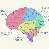
Researchers from the University of California, Los Angeles examined 16 adult brains using post mortem scans of the temporal lobes, specifically the hippocampus. The researchers wanted to determine shrinkage, or atrophy, or the region using seven Tesla MRI scanners for 60 hours each. Nine of the brains were from Alzheimer’s disease patients and the remaining ones were from cognitively normal participants. Memory function is a primary function of the hippocampus, the researchers explained, which can shed light on the earliest symptoms of Alzheimer’s disease, such as memory loss.
The researchers then analyzed the buildup of amyloid tau protein and loss of neurons, which indicated Alzheimer’s disease development. The researchers said these scans allowed the researchers unprecedented visualization of the hippocampal tissue.
The brain scans showed the protocol for measuring the hippocampus was successful. The MRI scans aligned with the pathological changes that were already observed in the patients’ brains and were noted hallmarks of Alzheimer’s disease: progressive development of amyloid plaques and neurofibrillary tangles in the brain.
“This hippocampal protocol will now become the gold standard in the field, adopted by many if not all research groups across the globe in their study of Alzheimer’s disease,” study leader Liana Apostolova said in a press release. “It will serve as a powerful tool in clinical trials for measuring the efficacy of new drugs in slowing or halting disease progression.”
Previous methods to measure the hippocampus have not been as successful or valid. Typically, the press release explained, the volume of a hippocampus is about 3,000 to 4,000 cubic millimeters. But a pair of scientists could each come up with a difference nearing 2,000 cubic millimeters while measuring the same brain area. There have also not been studies verifying the estimates for volume of the hippocampus using MRI scans to correspond to actual tissue loss.
To conduct this research and address these faulty measurements, the investigators created a Harmonized Protocol for Hippocampal Segmentation (HarP) which outlines a definitive method for measuring hippocampal atrophy through structural MRI. This method, they said, best corresponds to the Alzheimer’s disease onset process.
“The technique is meant to be used on scans of living human subjects, so it’s important that we are absolutely certain that this methodology measures what it is supposed to and captures disease presence accurately,” Apostolova concluded. “As a result of the years of scientifically rigorous work of this consortium, hippocampal atrophy can finally be reliably and reproducibly established from structural MRI scans.”