Leg Lesion in an Older Woman
This 71-year-old woman presented with an itchy raised erythematous leg lesion that had been present for 10 months. She has a history of significant sun exposure in the past and was prescribed topical triamcinolone 4 months ago which she believes has made it slightly smaller and less itchy.
Erythematous Leg Lesion in an Older Woman
This 71-year-old woman presented with an itchy raised erythematous leg lesion that had been present for 10 months. She has a history of significant sun exposure in the past and was prescribed topical triamcinolone 4 months ago which she believes has made it slightly smaller and less itchy.
What is your diagnosis?
- Squamous cell carcinoma
- Keratoacanthoma
- Prurigo nodularis
- Amelanotic melanoma
- Common wart
Diagnosis

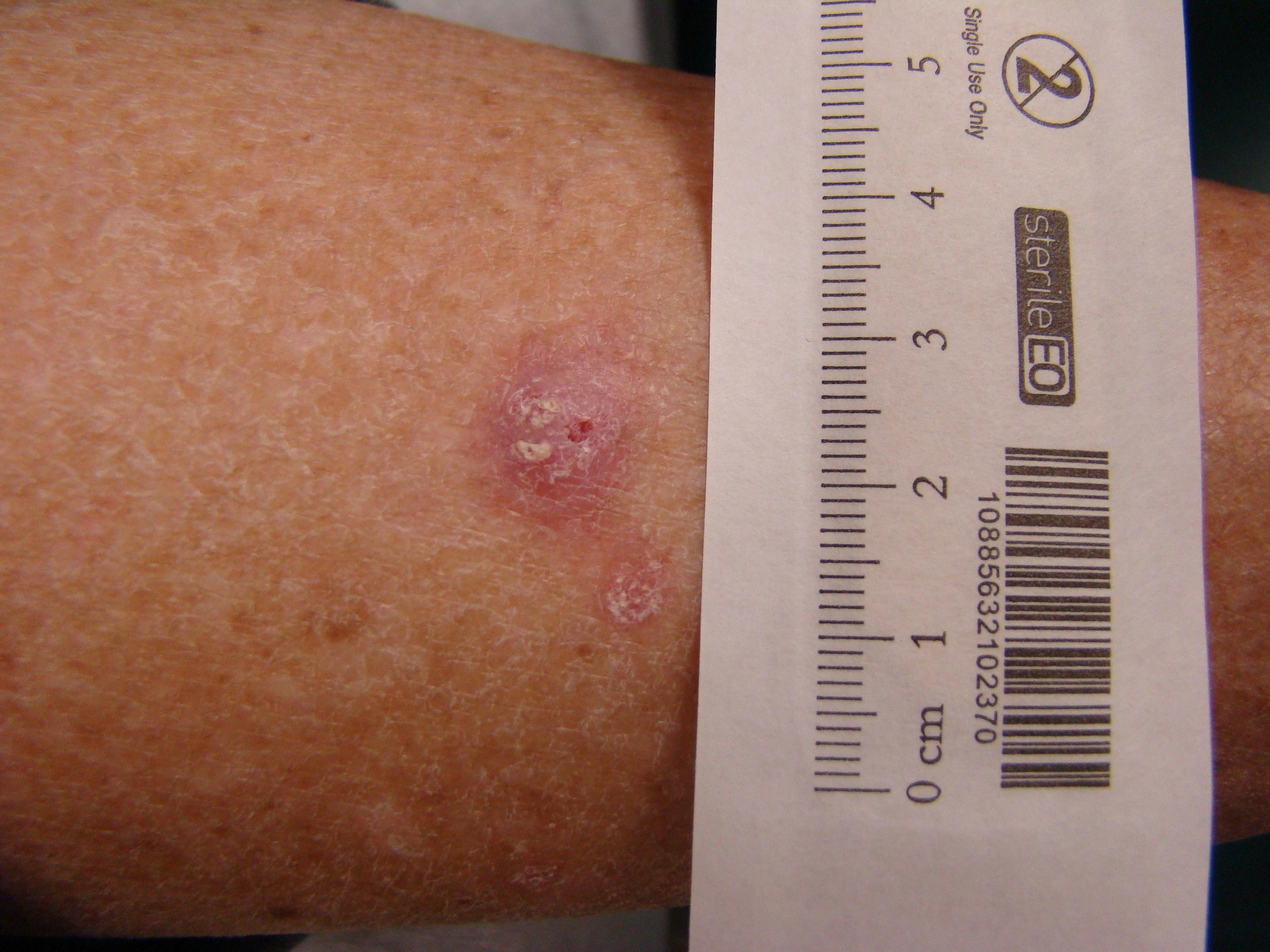
A deep shave excision confirmed this woman’s keratoacanthoma with clear margins. Although this lesion lacks the classic central keratin plug, the relatively large size (approximately 1 cm,) short duration and raised dome shape pointed to the diagnosis. Keratoacanthomas are considered by some to be a relatively indolent form of squamous cell carcinoma. Others classify it as benign since most will spontaneously resolve over time.
Unfortunately they tend to heal with significant scarring and with the difficulty in differentiating them from squamous cell carcinoma even with partial biopsy. The recommended treatment is complete excision. (1)
Squamous cell carcinoma (SCC) should enter the differential diagnosis of new erythematous lesions on sun-exposed skin in older patients especially with light-colored skin. There is often associated actinic change as actinic keratosis are precursor lesions to SCC and the lesions tend to be more slowly growing versus keratoacanthomas which enlarge and become thick rather quickly versus lateral spread.
Prurigo nodularis would also present as an itching raised lesion, but would typically be more excoriated and lichenified due to repeated scratching. Multiple lesions may also be present along with a history of anxiety or picking at the skin lesions. (2)
Melanoma, including amelanotic (non-pigmented) melanomas should be considered in leg lesions especially with female patients. These lesions often erode and bleed by the time they reach this size and are typically more irregular. Pathology is important to confirm the diagnosis. Dermoscopy may reveal ulceration, atypical vessels and whitish streaks. (3)
Common warts also called verruca vulgaris can also present as domed shaped papules (less than 5mm) or even nodules (greater than 5mm.) In contrast to this patient, warts which are due to human papilloma virus, are more common in younger individuals (4), the lesions are often multiple, and they usually have a rough irregular surface.
About the Author


Daniel Stulberg, MD, is a Professor of Family and Community Medicine at the University of New Mexico. After completing his training at the University of Michigan, he worked in private practice in rural Arizona before moving into full-time teaching. Stulberg has published multiple articles and presented at many national conferences regarding skin care and treatment. He continues to practice the full spectrum of family medicine with an emphasis on dermatology and procedures.
References
2. Ustine, RP, Stulberg, DL, Colver, GB Cutaneous Cryosurgery 4th edition CRC press 2014
3. Giacomel J, Zalaudek I. Pink lesions. Dermatol Clin. 2013 Oct;31(4):649-78.