Pros and Cons of Echocardiography Technologies in Diagnosing Stress Cardiomyopathy
Stress cardiomyopathy is a unique cardiac syndrome in which transient left ventricular (LV) systolic dysfunction mimics acute myocardial infarction (AMI). It is usually brought on by acute emotional or physical stress (or both) and has 3 distinctive features: acute LV wall dysfunction, absence of significant obstructive coronary artery disease, and rapid improvement of LV systolic function within days or weeks.


Stress cardiomyopathy is a unique cardiac syndrome in which transient left ventricular (LV) systolic dysfunction mimics acute myocardial infarction (AMI). It is usually brought on by acute emotional or physical stress (or both) and has 3 distinctive features: acute LV wall dysfunction, absence of significant obstructive coronary artery disease, and rapid improvement of LV systolic function within days or weeks.
It has many names—Takotsuba cardiomyopathy(TCC), LV apical ballooning syndrome, and broken heart syndrome.
The likely cause, Rodolfo Citro, MD, PhD and colleagues report in the Journal of the American Society of Echocardiography is the heart’s response to a surge of catecholamines triggered by a stressful event.
It is more common in women, with 90% of incidences occurring in women ages 56 to 90 and the rest, mostly in men of the same age range. Fewer than 10% of cases are in people under age 50.
These patients are generally thought to recover quickly, within a few days or weeks. But newer studies suggest “both short- and long-term mortality may be substantial an similar to that of patients with acute coronary syndromes,” Citro writes.
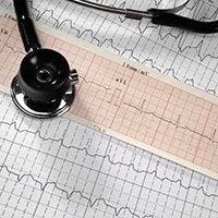
The fastest and least invasive way to diagnose the condition is with echocardiography (ECG)—both standard and advanced.
The authors evaluate the options available with advanced ECG and list its advantages and disadvantages.
Among the pluses, advanced ECG has to potential to offer a more detailed analysis of regional and global myocardial contractility by using 2D speckle tracking. But it’s a sophisticated technology requiring an expert operator and is difficult to perform in emergency settings, Citro concludes.
Strain Doppler, another feature of advanced ECG, can determine the degree of myocardial stunning, but is difficult to recognize in patients with arrhythmias. Contrast ECG, another advanced technique, offers a more precise definition of ejection fraction, but requires using several contrast agents that are not good for acutely ill patients. Vasodilator stress ECG, similarly requires use of dipyiradome and adenosine with should be used with caution in critically ill patients.
In all, the authors evaluate 7 such techniques and found none without drawbacks.
Still, they concluded advanced techniques are providing “further mechanistic and pathophysiologic insights.”
Standard ECG is less sophisticated but can also identify important distinctive features of TTC.
Whichever method is used “prompt imaging of ventricular function is the key to making the diagnosis,” he writes.