Bilateral Hand Pain in a Patient With Chronic Hepatitis C and Sarcoidosis
A 51-year-old African American man with a 15-year history of chronic hepatitis C and sarcoidosis presented with persistent bilateral hand pain. The pain was aggravated by physical activity and had a waxing-and-waning pattern.
A 51-year-old African American man with a 15-year history of chronic hepatitis C and sarcoidosis presented with persistent bilateral hand pain. The pain was aggravated by physical activity and had a waxing-and-waning pattern.
The man had an inadequate response to therapy, which included corticosteroids, methotrexate
(MTX), and hydroxychloroquine (HCQ). There was no associated rash, numbness, tingling, or weight loss.
On examination, both hands were noted to have swollen digits with bony enlargements of the proximal interphalangeal and metacarpophalangeal joints. The left index and middle fingers had
mild swan-neck deformities with signs of mild synovitis. There were no skin nodules.
Bilateral muscle strength and handgrip were intact. Bone biopsies of the patient’s right tibia, great toe, and medial malleolus were performed because there was pain and swelling in the right ankle.
X-ray films of both of his hands were obtained and are seen below.
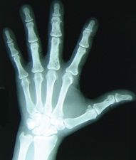
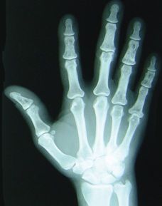
What do they show? What is your diagnosis?
(Find the answer on the next page.)
The clinical and radiographic findings suggested that the patient had sarcoid arthritis.
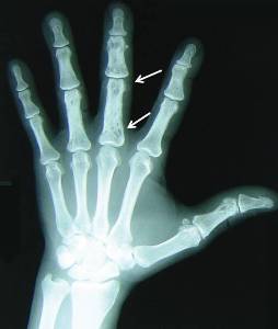
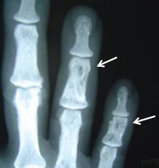
The hand x-ray films showed bilateral multiple lucencies in the proximal, middle, and distal phalanges consistent with bone sarcoidosis. Closer views showed cystic changes in the proximal and middle phalanges, with a lack of periarticular osteopenia or erosions. Specifically, large intraosseous cystic areas were seen in the middle third phalanx in the patient’s left hand (arrows, left), and intraosseous cysts were seen in the middle phalanges of the fingers of the right hand (arrows, below right).
Sarcoidosis is a multisystem disorder characterized by noncaseating granulomas in various affected organs. Typically, patients present with bilateral hilar adenopathy, pulmonary infiltrates, and skin or eye lesions.
Rheumatologic manifestations are uncommon but may be significant in acute disease (4% to 38%), involving the joints, bones, periarticular soft tissue, and muscles.1 About 15% to 25% of patients with sarcoidosis present with either acute or chronic arthritis. Acute sarcoid arthritis may present as joint pain that involves the ankles, knees, wrists, and elbows and, less frequently, the small joints of the hands and feet.The disease usually is self-limited and has a favorable outcome.
Chronic sarcoid arthritis is rare and has been seen mainly in African American patients. It is associated with long-standing sarcoidosis and may be persistent, with periods of remission and exacerbation.2 Usually, it is polyarticular and involves the shoulders, hands, wrists, ankles, and knees. Dactylitis similar to that seen in psoriatic arthritis and Jaccoud-type deforming arthropathy also has been described.
Osseous involvement occurs in 5% of patients with sarcoid arthropathy and usually is asymptomatic. It is seen in long-standing sarcoidosis with multiorgan involvement, especially infiltrative skin disease. Osseous involvement causes cystic, reticular, or sclerotic lesions-mainly of hands and feet-with lesions of the heads of the proximal and middle phalanges and, less frequently, the metacarpals. Punched-out cysts may be seen on radiographs, along with tunneling, remodeling, and destruction of the cortex, which may lead to multiple fractures and sequestrum formation.
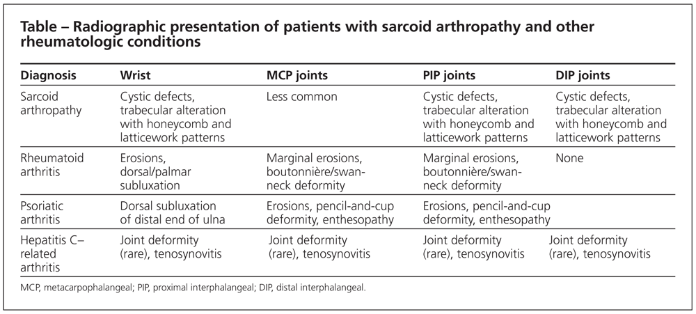
The differential diagnosis in this patient included rheumatoid arthritis, psoriatic arthritis, lymphoma,
and hepatitis C–related arthritis; the various conditions present differently on radiographs (Table).The patient had symmetric polyarthralgia resulting from sarcoid arthropathy and osseous sarcoid. The bone biopsy of the right tibia, great toe, and medial malleolus revealed noncaseating granulomas.
In this case, the arthropathy represents a chronic manifestation of sarcoid arthropathy.The patient did not have skin lesions or a history of erythema nodosum; he does have sarcoid lung disease and had elevated liver function test results because of chronic hepatitis C.
NSAIDs are the initial therapy for patients with acute sarcoid arthritis. Patients with chronic sarcoid
arthritis often also require corticosteroids. In others, colchicine or HCQ may be used. MTX may be an alternative in corticosteroid-refractory cases.There are no conclusive data about the efficacy of biologic agents, such as the tumor necrosis factor _ inhibitors. Natural progression of the lesions may lead to destruction of the involved bone and subsequent deformities. In a few cases, it can cause cord compression in the spine. The response to treatment is generally poor.
Initially, our patient was treated with NSAIDs; later, he received corticosteroids, HCQ, and MTX. Prednisone was started in 2000; the patient’s current dosage is 15 mg/d. MTX was started in 2003; the current dosage is 12.5 mg/wk. HCQ was started in 2002; the current dosage is 400 mg/d. All these medications are being given for pulmonary sarcoidosis. In addition, the patient needed low-dose narcotics for pain control. He received no treatment for hepatitis C.
This case was submitted by Ramesh Pappu, MD, associate professor of medicine and rheumatology fellowship program director,and Humaira Hussain, MD, rheumatology fellow, at Drexel University College of Medicine in Philadelphia.
References:
1. Visser H, Vos K, Zanelli E, et al. Sarcoid arthritis: clinical characteristics, diagnostic aspects, and risk factors. Ann Rheum Dis. 2002;61:499-504.2. Abril A, Cohen MD. Rheumatological manifestations of sarcoidosis. Curr Opin Rheumatol. 2004;16:51-55.