Uniform Brain Patterns May Provide Better Understanding of Severe Mental Illnesses
Providing additional insight into mental illnesses such as schizophrenia and bipolar disorder, recent research has found neurological operations at rest are similar to those seen during active tasks.


Providing additional insight into mental illnesses such as schizophrenia and bipolar disorder, recent research has found neurological operations at rest are similar to those seen during active tasks.
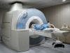
In the July 2, 2014, issue of Neuron, researchers at Rutgers University-Newark and Washington University in St. Louis utilized functional magnetic resonance imaging (fMRI) in participants who conducted 64 tasks, which were “deï¬ned as distinct cognitive processes such that the same stimuli could be presented across each of the 64 tasks, but distinct cognitive processes would be necessary to respond correctly to each one,” the study authors wrote.
Additionally, the investigators added 7 tasks to the study from the Human Connectome Project dataset comprised of highly varying activity that tapped into different brain facilities. In doing so, they reportedly discovered a “whole-brain network architecture present across dozens of task states that was highly similar to the resting-state network architecture.”
Since frequent functional connectivity strengths were similar during resting and active tasks, the results suggest that functional brain organization is intrinsic in nature, the authors said, noting their findings create a new relationship between resting-state functional connectivity and task-evoked functional connectivity, which were formerly viewed as separate entities by neuroscientists.
“These results indicate the brain’s functional network architecture during task performance is shaped primarily by an intrinsic network architecture that is also present during rest, and secondarily by evoked task-general and task-specific network changes,” the researchers wrote.
Furthermore, the investigators reported that their study “revealed a complex set of functional connectivity changes, with a prominent pattern of decreased within-system functional connectivity during task performance,” though they expressed future research will be needed to clarify the meaning behind the trend and why it occurs during tasks irrespective of activity status.
In a statement provided by Rutgers University-Newark, study contributor Michael Cole, of the Center for Molecular and Behavioral Neuroscience, commented that the findings were a relief, since brain organization was thought to change during every task.
“We can now observe people relaxing in the scanner and be confident that what we see is there all the time,” Cole stated. “If that had (not) been the case, we would have had less hope that we could understand mental illness in our lifetime.”
Thus, the investigators concluded that their research “provides a framework for future studies to characterize functional connectivity during tasks, in terms of both intrinsic functional connectivity identiï¬ed using resting states and evoked functional connectivity identiï¬ed across tasks and in particular task contexts.”