Diagnosis and Management of Gout in 2011
Early and accurate gout diagnosis and disease management are essential. Making the clinical diagnosis takes into consideration the differential diagnosis supported by the use of clinical, serological, and diagnostic studies.
ABSTRACT: Early and accurate gout diagnosis and disease management are essential. Making the clinical diagnosis takes into consideration the differential diagnosis supported by the use of clinical, serological, and diagnostic studies. Hyperuricemia is a serum uric acid level consistently higher than 6.8 mg/dL. Various imaging modalities have advantages over others in making the diagnosis. The impact of gout is underestimated, and diagnosis and management generally are poor. Everyday challenges include medication noncompliance, nonadherence, and nonconcordance. The contemporary approach to gout management is focused on controlling 3 distinctive phases of disease: acute flare, flare prophylaxis, and reduction of the body urate burden.
______________________________________________________________________________________
Diagnosis
Clinical features of the acute gout attack
Help from the laboratory
Imaging studies
Management
Three phases of disease
Hyperuricemia and gout have increased markedly in prevalence and clinical complexity over the past 2 decades. Early and accurate diagnosis and disease management are essential, because appropriate management limits systemic disease involvement, health care costs, unnecessary hospitalizations, workdays lost, and functional disability.
Making the clinical diagnosis of gout takes into consideration the differential diagnosis supported by the use of clinical, serological, and diagnostic studies. As with all diagnostic evaluations, the history and physical examination direct the process.
In this article, we describe the history and physical examination, the clinical features of an acute gout attack, laboratory testing, appropriate imaging studies, approaches to treatment, and various agents that may be effective. Included are recommendations for specialized management and referral to a rheumatologist for help in management.
DIAGNOSIS
History and physical examination
Understanding the predominance of gout in men is important for a complete evaluation (ratio, 20:1). The onset of gout in men usually is seen between age 35 and 50 years. In women, gout is seen largely after menopause; estrogen is uricosuric and plays a role in controlling urate levels.
Understanding patterns in affected joints also is important. The development of microtophi in cooler synovium and in the cartilage of distal extremity joints, olecranon bursa, and helix of the ear explains the predilection of lower extremities for gout flare. Urate is less soluble at temperatures below 37°C (98.6°F). Deeper, warmer structures become affected when the urate burden is great and over a long period. Urate precipitation also is favored by the absolute degree of supersaturation, plasma proteins, and abnormal aggrecans (eg, osteoarthritis [OA]), which reduce solubility.
Food and alcohol consumption both trigger acute flares and contribute to long-term management issues. Gout incidence is associated with daily intake of red meat and seafood, which have a high purine content, as well as consumption of alcohol, especially beer, which has the highest soluble purine content. High-purine-content vegetables did not seem to impact incidence of gout.1 There is a direct enzymatic link between increased intake of fructose (high-fructose corn syrup in artificial sweeteners) and elevated serum urate level2-a concern because fructose is ever more predominant in food. This links directly to the rise in obesity, a common denominator in the comorbidities of hyperuricemia, hypertension, hyperlipidemia, and atherosclerosis.
Increasing data indicate that gout and hyperuricemia alone are associated with increased cardiovascular disease (CVD) and mortality.3,4 Some authors argue that hyperuricemia is a biomarker for increased CVD, hypertension, and new-onset and progressive renal disease. Therefore, these comorbidities and the medications currently used to manage them are important to consider-they complicate the diagnosis as well as the management of acute, chronic, and recurrent gouty arthritis. Insufficient urate excretion can be iatrogenic with widely used therapies, such as diuretics, low-dose aspirin, niacin, cyclosporine, tacrolimus, antituberculosis drugs, and many antivirals.
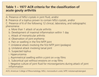
TABLE 1

1977 ACR criteria for the classification of acute gouty arthritis
Clinical features of the acute gout attack
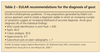
The current diagnostic criteria for acute gouty arthritis are based on the 1977 American College of Rheumatology (ACR) criteria (Table 1). The ACR criteria for the classification of acute gout are inadequate for in-office diagnosis in about 20% of cases.5 New criteria currently are under development. European League Against Rheumatism (EULAR) recommendations for the diagnosis of gout highlight the value of synovial fluid crystal analysis (Table 2).
However, the classic description of acute gouty arthropathy stands: It is the very rapid onset of unilateral monarthritis with acute inflammation and severe pain. Often called podagra, it usually involves the midfoot or the first metatarsophalangeal joint. Patients who have this symptom complex often report previous episodes of the same symptoms. At first, these attacks are self-limited, resolving in 3 to 10 days. Also associated with classic acute gouty arthritis are periarticular or bursal erythema, followed by skin desquamation with resolution of attack as well as systemic symptoms, such as fever and leukocytosis. Uncharacteristic polyarticular involvement also may occur but often in the later stages of the disease.
These features can make the diagnosis difficult because similar signs and symptoms are seen in other acute processes, including septic arthritis, calcium pyrophosphate dihydrate (CPPD) crystal deposition disease, OA, posttraumatic arthritis, and soft tissue injury.
TABLE 2

EULAR recommendations for the diagnosis of gout
Help from the laboratory
Hyperuricemia is a serum uric acid (SUA) level consistently higher than 6.8 mg/dL. Note that during an acute gout flare, levels may be normal or only slightly elevated (lower than 8 mg/dL),6 often leading clinicians to a misdiagnosis and, ultimately, mismanagement. Also note that asymptomatic hyperuricemia is common and does not need to be managed unless there is systemic or joint involvement.
Because of these confounders, a joint aspiration must be completed for synovial fluid analysis if the diagnosis is at all indeterminate because it remains the gold standard for the diagnosis of gout. Musculoskeletal ultrasonography (MSUS) and dual energy CT (DECT) are beginning to challenge this standard-they are becoming useful alternative, noninvasive means to assess patients for the changes of acute, intermediate, and chronic gout.
No current diagnostic study can effectively differentiate between infectious and noninfectious causes of inflammatory arthritis other than aspiration and fluid analysis with cell count, Gram stain, and cultures. These are imperative for the most accurate diagnosis and directed management. The EULAR recommendations for the diagnosis highlight the value of synovial fluid analysis. Joint fluid is assessed with the use of a polarized microscope looking for negatively birefringent needle-shaped crystals.
During an acute gout flare, the joint aspirate is inflammatory. A useful rule of thumb when grossly evaluating synovial fluid is the inability to read print on paper through the fluid-filled tube-this would indicate aspirate with a leukocyte count into the inflammatory range of 1000 to 2000/µL. Aspirates with leukocyte counts higher than 1000 to 2000/µL are very cloudy. In an attempt to differentiate between gout and bacterial/septic arthritis, a leukocyte count with differential is just as important as Gram stain and culture. In gouty arthritis, the white blood cell (WBC) count typically is between 1000 and 50,000/µL, with less than 90% neutrophils; in septic arthritis, the WBC count typically is greater than 50,000/µL, with more than 90% neutrophils.
There is a direct inverse relationship between the amount of inflammation within the joint and the viscosity of the joint fluid. Crystal analysis is completed by assessment under a polarized light microscope, which requires only a minute amount of fluid.
Monosodium urate (MSU) crystals, which are precipitated in gout, are needle-shaped and strongly negative birefringent, meaning that they appear yellow when the long axis of the crystal is parallel to the red compensator and blue when it is perpendicular. In contrast, CPPD crystals have various shapes, although they primarily are rhomboid-shaped with weak positive birefringence. Additional crystal formations may be seen.
Previous corticosteroid injections may confuse the diagnosis because they appear as strongly positive birefringent crystals. During an acute attack, MSU crystals are seen both extracellularly and intracellularly as they are engulfed by the leukocyte (neutrophil). The fluid should be analyzed for at least 5 minutes over numerous fields to firmly rule out the presence of MSU crystals.7 For correct visualization of the aspirate, the handling of the sample is extremely important.
Imaging studies
Plain radiography. This study demonstrates nonspecific soft tissue changes of swelling as well as more definitive changes characteristic of gout, including subcortical cysts, marginal “punched-out” erosions with overhanging edges (new cortical bone formation), and calcified tophi. The advantages are low cost, quickness, low radiation dose, in-office examination, and widespread availability. The disadvantages include a lack of sensitivity and specificity in early disease. After 5 years of flare activity, only about half of patients with gout have radiographic or erosive changes on plain radiographs.7,8
High-resolution CT. The advantages are superiority to plain radiography for detecting early disease and tophi changes. The disadvantages are high cost, high radiation exposure, moderate availability, and a lack of specificity.
DECT. The advantages of this newer diagnostic study are sensitivity and specificity for urate deposits, especially those in soft tissue and bone structures.9 The disadvantages: expensive and high radiation exposure.
MRI. This modality detects tophi with representative decreased signal in both T1- and T2-weighted images with variable enhancement.8 MRI is a useful examination when the presence of tophi is suspected but not proven. MRI often demonstrates greater-sized tophi than expected or appreciated on physical examination. The advantages are superiority to plain radiography in early detection and characterization of tophi and no radiation exposure. The disadvantages are less benefit than CT scanning, a lack of specificity for tophi, moderate availability, and high expense.
High-resolution (8 to16 MHz) MSUS. This can be a point of care technology for a quick and accurate bedside evaluation. It is sensitive for early detection of crystal deposits, seen as hyperechoic masses or linear bands in synovial or cartilage borders8 with the characteristic “double-contour” sign (representing MSU crystals layering the articular cartilage surface).
MSUS can differentiate between MSU crystal deposits and CPPD crystal deposits and is far more sensitive than plain radiography for early disease detection.8 The advantages are sensitivity and specificity, good early disease detection, low cost, widespread availability, portability, in-office use, and no radiation exposure. Because MSUS often is available for in-office services and does not use radiation, with a lower cost than CT and MRI, it is a popular choice with both patients and physicians.10 A disadvantage: a sonographer skilled in musculoskeletal evaluations and interpretation is required.
MANAGEMENT
The impact of gout is underestimated, and diagnosis and management generally are poor. Misunderstanding of the treatment objectives by patients and all too often by clinicians caring for them is widespread. A lack of consensus on many aspects of gout care, ineffective communication, and even argument among providers comes from a lack of guidelines and poor implementation of those that are available. Old-school thinking abounds, especially about the duration of both urate-lowering therapy and medications for acute gout flares.
Everyday challenges include medication noncompliance (failure or refusal to take prescribed medications), nonadherence (failure to take prescribed medications correctly), and nonconcordance (failure to agree on the therapeutic plan). Another challenge is the limitations of the medications used to manage gout because of comorbidities, drug-drug interactions, and advanced progression of the disease process.
Of the 8.3 million diagnoses of gout in the United States, urate-lowering therapy (ULT) was started for only 60%. Of those 5 million patients, only 0.5 million achieved the target SUA level of lower than 6 mg/dL; 4.5 million are considered “treatment failures” because of noncompliance (0.55 million), nonadherence (3.2 million), and nonconcordance (0.76 million). Of the 4.5 million treatment failures, only 18% were considered to have truly “difficult-to-treat” gout.11-13
Patient/provider issues resulted in treatment failure in 83% of patients for whom ULT was started. Patient issues included the well-known error of confusing therapies for managing acute gout symptoms with those that lower the urate burden. Many patients have had very bad experiences with previous therapies. In addition, they cling to past inappropriate medical advice, do not understand the treatment course, become impatient, and avoid healthy lifestyle changes. Provider-associated challenges include lacking unawareness of the need for a “treat-to-target” approach, no monitoring of the SUA level as a biomarker of disease, and seeing gout as only an acute process.
Three phases of disease
The contemporary approach to gout management is focused on controlling the following 3 distinctive phases of disease:
Acute flare. The goals of therapeutic management are to provide rapid analgesia for the flare and inhibition of acute inflammation to provide a swift return of pain-free function in involved joints in a safe and cost-efficient manner. During an acute flare, the urate crystals that have deposited along the joint surface in the form of asymptomatic microtophi are, for reasons under investigation, released into the joint fluid, exposing themselves to the resident cells within the joint (synovial lining, mast cells, and macrophages). This results in activation of leukocytes, primarily neutrophils and monocytes, which act to propagate and prolong the inflammatory cascade. If left unmanaged, this inflammatory phase would be self-limited in about 3 to 10 days for patients with early disease and 10 to 20 days in patients with late disease.
With effective pharmacological intervention, a patient can expect 50% improvement in pain within 2 or 3 days.14 Rapid analgesia and inhibition of the inflammatory process are directly correlated with the time interval between the onset of flare/attack and the start of anti-inflammatory therapy. The sooner the better: therapies started after 72 hours have little impact on the course of the flare.
Medication targets for the management of an acute gout flare are suppressing the expression, secretion, and signaling of inflammatory cytokines. They are the key players, affecting neutrophil adhesion, migration, and activation. Blunting their production results in decreased neutrophil recruitment into the joint space and a limited inflammatory response.
Current first-line pharmaceutical options include NSAIDs and cyclooxygenase 2 inhibitors, oral low-dose corticosteroids, and low-dose colchicine. The therapeutic action of colchicine is slightly different in that it targets and binds tightly to tubulin, which inhibits microtubule elongation and function within the phagocytes and endothelium. Colchicine given at low doses suppresses neutrophil adhesion and migration; high doses inhibit inflammatory mediator release.
Second-line options include corticosteroids (intramuscular, intravenous, intra-articular), off-label adrenocorticotropin hormone, and off-label interleukin (IL)-1 inhibitors. IL-1 inhibitors, anakinra, and canakinumab are emerging regimens for the management of acute attacks.
In a small pilot study, 10 patients for whom other anti-inflammatory therapies for acute gout were not successful were treated with anakinra.15 All patients responded rapidly within 24 hours and reported rapid, significant symptom relief within 48 hours of initial injection and no adverse effects.
The effectiveness of the IL-1 inhibitor canakinumab, 150 mg given subcutaneously, was compared with that of the corticosteroid triamcinolone, 40 mg given intramuscularly, in patients who had contraindications for NSAIDs or colchicine.16 Primary outcome measures were at 72 hours post-dose. Canakinumab proved to be superior to triamcinolone.
Currently there is no standard treatment for patients with acute gout flare. Selection of the intervention should be individualized, taking into account the patient’s allergies and comorbidities.
Long-term management of gout is effective only if it involves a comprehensive plan with flare prophylaxis strategies that not only incorporate medication intervention but also trigger identification. Such a plan often requires modification of diet and alcohol intake as well as agreement between the patient and physician about goals and responsibilities. ULT is at the core of an effective long-term management scheme of reducing the underlying metabolic cause of gout. Because perturbation in uric acid levels often predicates gout flares, gout attack prophylaxis must be understood and used while ULT is started and escalated.
Flare prophylaxis. Strategies are based on medications that control the rapid development of inflammation that occurs when microtophi or tophi are destabilized within the joint. Gout attack prophylaxis encompasses the time period from second attack through the start and escalation of ULT until at least 3 months after target SUA levels are reached.
Acute flares with ULT, termed “gout mobilization” flares, are associated with shifts in SUA levels that occur during the start of these therapies and are noted in dosage changes. Best-practice guidelines recommend low-dosage colchicine, 0.6 to 1.2 mg/d, as first-line therapy. Low-dosage NSAIDs (eg, naproxen, 250 mg bid, or indomethacin, 25 mg bid) are second-line treatment if the patient has contraindications to colchicine (eg, severe renal disease). Prophylaxis should continue for at least 3 months after ULT normalizes SUA levels to 6 mg/dL or lower. Prednisone is a poor choice for gout prophylaxis.
Colchicine acts by suppressing both the baseline level of subclinical inflammation in the gouty joint and the augmentation of inflammation that may come with flare.17 Adverse effects of cholchicine are dose-related. Most are GI complaints that can be managed by decreasing the dose.
Patient and physician education that includes dietary trigger modifications is an important element in both gout flare prophylaxis and long-term gout management. Education must be manageable and aimed at behavioral changes in exercise, diet/alcohol intake, and treatment adherence, along with awareness of what the overall management plan is.
Patients should know what their current SUA level is and their target goal. Just as in other chronic diseases, such as hypertension and diabetes mellitus, numbers matter. A basic understanding of disease pathophysiology can be made complex or simple to meet an individual patient’s need. Emphasizing the importance of continuing the treatment regimen-specifically not to discontinue or change ULT with an acute attack-is critical to success. Having adequate amounts of medication to both manage flares and continue daily ULT is essential.
Newer therapies to rapidly abort gout flare and prevent flare during ULT initiation are in development. Rilonacept currently is in phase 3 trials for prevention of gout flares during the start of ULT.18 This IL-1–blocking formulation was tested with 80- and 160-mg subcutaneous weekly injections versus placebo for 16 weeks. Rilonacept showed a marked reduction in acute flares during the first 16 weeks after ULT initiation and dose escalation, with adverse effects similar to those with placebo.
Long-term management of gouty arthritis includes dietary and educational recommendations, with the additional use of ULT. The xanthine oxidase inhibitors allopurinol and, more recently, febuxostat are first-line treatments. For add-on or second-line therapy, probenecid-a uricosuric agent-is used occasionally, but it has limits. If an estimated glomerular filtration rate (eGFR) lower than 50 mL/min, a history of kidney stones, or a state of uric acid overproduction is established as the cause of the hyperuricemia, probenecid is contraindicated.
With oral agents, it may take years for patients with refractory chronic tophaceous gouty arthritis to reach therapeutic goals. Functional impairment in these patients can be considerable and result in poor quality of life.
Pegloticase, an infusible recombinant uricase, was FDA-approved in 2010 for use in patients whose gout is recalcitrant to conventional therapies or who have large tophaceous deposits. It can suppress SUA levels rapidly and accelerate tophus resorption.
Reduction of the body urate burden. The goal of all ULT is to prevent disease progression by reducing the body urate burden. The only successful approach to preventing flares and lessening the destructive potential of gout is to bring the SUA level well below the solubility threshold of 6.8 mg/dL and to reduce the total body urate burden. The indications for starting ULT are more than 1 flare per year, advanced gouty arthritis, the presence of tophi, a uric acid overproducing state, daily urinary uric acid excretion greater than 1000 mg, recurring renal stones, gout with chronic renal disease, and gout with major organ transplant.
Treat-to-target is the strategy used to achieve superior outcomes in eliminating gout flares, resolving tophi, and reducing the impact of chronic gouty arthritis, thereby reducing disability and pain with improved quality of life. Once the evidence-based SUA target level of lower than 6 mg/dL has been achieved, the objective is to maintain the level lifelong, which requires monitoring every 6 months. Both patients and physicians must be committed to this goal.
Allopurinol has been a widely used ULT over the past 4 decades. FDA approval is for dosages from 100 to 800 mg/d. However, primary care physicians seldom exceed dosages of 300 mg/d. At that dosage, only 40% of patients with gout reach the target SUA goal.19 The reason is complex, but largely it is the paucity of evidence relating to efficacy and safety for dosages exceeding 300 mg/d. Treating to target often requires dosages of greater than 300 mg/d. When the 300-mg dose is exceeded, allopurinol is better tolerated if given in divided doses twice daily.
Rash may develop in 2% of patients. Most cases are benign and dose-related.
Febuxostat is a nonpurine analogue uricostatic agent structurally different from allopurinol; there have been no reports of cross-reactive toxicities. Unlike with allopurinol, there currently is no recommendation for dose reduction in mild to moderate renal impairment.
In the CONFIRMS trial, 40 mg of febuxostat was similar to 300 mg of allopurinol in effectiveness, but 80 mg of febuxostat outperformed allopurinol.20 Like allopurinol, febuxostat or any drug can cause hypersensitivity. Febuxostat is not contraindicated in patients who have mild hypersensitivity reactions to allopurinol. Both of these medications can cause elevated transaminase levels and require ongoing monitoring.
Probenecid is the only approved uricosuric agent in the United States for patients who are urate underexcreters. Adequate renal function (eGFR higher than 50 mL/min) is required, and dosages from 250 to 1000 mg bid or tid are needed to reach target. Adequate hydration to prevent urolithiasis is important.
Benzbromarone is a potent uricosuric agent. However, its use is restricted and it is available only outside of the United States.
Pegloticase-the only FDA-approved uricolytic agent that dissolves the uric acid structure-is a PEGylated uricase enzyme that completes the final step in the purine catabolic pathway, creating soluble urate, which lacks the potential to precipitate. The uricase enzyme, through evolution, has disappeared in humans and higher primates, rendering them susceptible to the formation of microtophi and gout. The PEGylation of the uricase has prolonged its half-life and reduces immunogenicity, a problem for its predecessor recombinant uricase, rasburicase.
Pegloticase, 8 mg, is infused every 2 weeks. It is a biologic agent with the potential for immunogenicity best administered in the hands of those familiar with such infusions. The ability of pegloticase to produce and maintain very low SUA levels is impressive.21
Comprehensive gout control can be complex, including management in all 3 disease phases with additional emphasis on education and lifestyle risk reduction strategies.23 Medications should be patient-centered, incorporating all comorbidities and potential adverse effects as well as drug-drug interactions.
Several disease states can alter purine metabolism, including fraility in older patients, diabetes mellitus, chronic heart failure, chronic kidney disease, hemodialysis, organ transplant, and refractory gouty arthritis and hyperuricemia. Patients may require specialized management of gout because of the considerations of urate handling and pharmacological intervention. In such cases, consulting a rheumatologist for help is appropriate.
Problems/comments about this article? Please send feedback.
References:
References
1. Choi HK, Atkinson K, Karlson EW, et al. Purine-rich foods, dairy and protein intake, and the risk of gout in men. N Engl J Med. 2004;350:1093-1103.
2. Davies PM, Simmonds HA, Singer B, et al. Plasma uridine as well as uric acid is elevated following fructose loading. Adv Exp Med Biol. 1998;431:31-35.
3. Feig DI, Kang DH, Johnson RJ. Uric acid and cardiovascular risk [published correction appears in N Engl J Med. 2010;362:2235]. N Engl J Med. 2008;359:1811-1821.
4. Gaffo AL, Edwards NL, Saag KG. Gout. Hyperuricemia and cardiovascular disease: how strong is the evidence for a casual link? Arthritis Res Ther. 2009;11:240.
5. Malik A, Schumacher HR, Dinnella JE, Clayburne GM. Clinical diagnostic criteria for gout: comparison with the gold standard of synovial fluid crystal analysis. J Clin Rheumatol. 2009;15:22-24.
6. Schlesinger N, Norquist JM, Watson DJ. Serum urate during acute gout [published correction appears in J Rheumatol. 2009;36:1851]. J Rheumatol. 2009;36:1287-1289.
7. Schlesinger N. Diagnosis of gout. Minerva Med. 2007;98:759-767.
8. Perez-Ruiz F, Dalbeth N, Urresola A, et al. Imaging of gout: findings and utility. Arthritis Res Ther. 2009;11:232.
9. Choi HK, Al-Arfaj Am, Eftekhari A, et al. Dual energy computed tomography in tophaceous gout. Ann Rheum Dis. 2009;68:1609-1612.
10. American College of Rheumatology Musculoskeletal Ultrasound Task Force. Ultrasound in American rheumatology practice: report of the American College of Rheumatology musculoskeletal ultrasound task force. Arthritis Care Res (Hoboken). 2010;62:1206-1219.
11. Zhu Y, Pandya B, Choi H. Increasing gout prevalence in the US over the last two decades: the National Health and Nutrition Examination Survey (NHANES). In: Proceedings from American College of Rheumatology Annual Meeting; November 6 - 11, 2010; Atlanta. Abstract 2154.
12. Sarawate CA, Brewer KK, Yang W, et al. Gout medication treatment patterns and adherence to standards of care from a managed care perspective. Mayo Clin Proc. 2006;81:925-934.
13. Riedel AA, Nelson M, Joseph-Ridge N, et al. Compliance with allopurinol therapy among managed care enrollees with gout: a retrospective analysis of administrative claims. J Rheumatol. 2004;31:1575-1581.
14. Rubin BR, Burton R, Navarra S, et al. Efficacy and safety profile of treatment with etoricoxib 120 mg once daily compared with indomethacin 50 mg three times daily in acute gout: a randomized controlled trial. Arthritis Rheum. 2004;50:598-606.
15. So A, De Smedt T, Revaz S, Tschopp J. A pilot study of IL-1 inhibition by anakinra in acute gout. Arthritis Res Ther. 2007;9:R28.
16. So A, De Meulemeester M, Pikhlak A, et al. Canakinumab (ACZ885) relieves pain and controls inflammation rapidly in patients with difficult-to-treat gouty arthritis: comparison with triamcinolone acetonide. In: Proceedings from the American College of Rheumatology Annual Meeting; November 6 - 11, 2010; Atlanta. Abstract 145.
17. Terkeltaub RA. Colchicine update: 2008. Semin Arthritis Rheum. 2009;38:411-419.
18. Terkeltaub R, Schumacher HR, Saag KG, et al. Evaluation of rilonacept for prevention of gout flares during initiation of urate-lowering therapy: results of a phase 3, randomized, double-blind, placebo-controlled trial. In: Proceedings from the American College of Rheumatology Annual Meeting; November 6 - 11, 2010; Atlanta. Abstract 152.
19. Takada M, Okada H, Kotake T, et al. Appropriate dosing regimen of allopurinol in Japanese patients. J Clin Pharm Ther. 2005;30:407-412.
20. Becker MA, Schumacher HR, Espinoza LR, et al. The urate-lowering efficacy and safety of febuxostat in the treatment of the hyperuricemia of gout: the COMFIRMS trial. Arthritis Res Ther. 2010;12:R63.
21. Sundy JS, Becker MA, Baraf HS, et al; Pegloticase Phase 2 Study Investigators. Reduction of plasma urate levels following treatment with multiple doses of pegloticase (polyethylene glycol-conjugated uricase) in patients with treatment-failure gout: results of a phase II randomized study. Arthritis Rheum. 2008;58:2882-2891.
22. Zhang W, Doherty M, Pascual E, et al; EULAR Standing Committee for International Clinical Studies Including Therapeutics. EULAR evidence based recommendations for gout, part I: diagnosis. Report of a task force of the Standing Committee for International Clinical Studies Including Therapeutics (ESCISIT). Ann Rheum Dis. 2006;65:1301-1311.
23. Choi HK. A prescription for lifestyle change in patients with hyperuricemia and gout. Curr Opin Rheumatol. 2010;22:165-172.
Recommended reading
• Terkeltaub R, Edwards NL. Gout: Diagnosis and Management of Gouty Arthritis and Hyperuricemia. 2nd ed. West Islip, NY: Professional Communications, Inc; 2011. This is a key resource that provides the most up-to-date review of gout diagnosis and treatment.