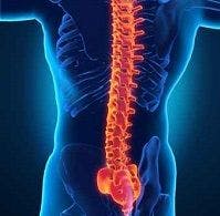
Fat Metaplasia Predictive of Spinal Progression in Spondyloarthritis
A study in PLOS One found evidence that fat metaplasia in sacroiliac joints (SIJ) is significantly associated with spinal progression for axial spondyloarthritis (axSpA).

A study in PLOS One found evidence that fat metaplasia in sacroiliac joints (SIJ) is significantly associated with spinal progression for axial spondyloarthritis (axSpA).
AxSpA falls into two categories: non-radiographic axSpA, in which there is no evidence of sacroiliitis on conventional radiographs, and ankylosing spondylitis (AS), in which there is definitive evidence of sacroiliitis. Radiographic progression in the spine is strongly associated with spinal mobility and functional status, and therefore represents a clinically important outcome and treatment target in those with axSpA.
Because spinal progression varies widely among patients with axSpA, it is clinically important to be able to predict radiographic spinal progression. Magnetic resonance imaging (MRI) can detect active inflammatory lesions in the SIJs, particularly on fat-suppressed (FS) images and can also detect both post-inflammatory changes including MRI-specific fatty lesions (fat metaplasia) and chronic changes (sclerosis, erosion, and ankyloses).
“…MRI of the SIJs is an important practical tool for evaluating patients suspected of having early spondyloarthritis,” the study authors wrote. This is the first study to examine the utility of inflammatory lesions on SIJ MRI for predicting spinal radiographic progression.
One hundred and ten patients who fulfilled the ASAS axSpA criteria were enrolled. All underwent SIJ MRI at baseline and lumbar spine radiographs at baseline, as well as after two years. Inflammatory and structural lesions on SIJ MRI were scored using the SPondyloArthritis Research Consortium of Canada (SPARCC) method. Spinal radiographs were scored using the Stoke AS Spinal Score (SASSS). Multivariate logistic regression analysis was performed to identify predictors of spinal progression.
Among the 110 patients, 25 (23%) showed significant radiographic progression (change of SASSS≥2) over two years. There was no change in the SASSS over two years according to the type of inflammatory lesion. Patients with fat metaplasia or ankyloses on baseline MRI showed a significantly higher SASSS at the conclusion than those without (p<0.001). According to univariate logistic regression analysis, age at diagnosis, the presence of fat metaplasia, erosion, and ankyloses on SIJ MRI and the presence of syndesmophytes at baseline, among other factors, were associated with spinal progression over two years. Multivariate analysis identified syndesmophytes and severe fat metaplasia on baseline SIJ MRI as predictive of spinal radiographic progression (OR, 14.74 and 5.66, respectively).
“The sequence of events that lead to the development of fat metaplasia in the SIJs is unclear,” the authors noted. “Little is known about the importance of this lesion, and its histopathology remains undefined. Fat metaplasia in the SIJs appears to be an inherent characteristic of the tissue response following the resolution of inflammation. Also, fat metaplasia is a key intermediary step in the development of SIJ ankylosis. The results presented herein suggest that fat metaplasia in the SIJs is associated with spinal progression.”
Limitations of the study include small sample size and the fact that the SASSS system takes into account the structural changes in the lumbar spine, but not those in the cervical and/or thoracic spines. The study also did not evaluate the effect of smoking on spinal progression due to insufficient baseline data.


