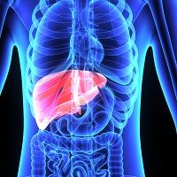
How Liver Damage Happens in NAFLD
In patients with non-alcoholic fatty liver disease, fat tissue insulin resistance can stimulate the activation of hepatic macrophages, due to an increased flux of free fatty acids. That results in liver damage, Italian researchers report.

In patients with non-alcoholic fatty liver disease, fat tissue insulin resistance can stimulate the activation of hepatic macrophages, due to an increased flux of free fatty acids (FFAs).
That causes liver damage, a team from Italy reports in an abstract to be presented at the International Liver Congress in Barcelona.
NAFLD has a bidirectional relationship with insulin resistance (IR): the liver is the target of an increased flux of FFAs and adipokines stemming from a dysfunctional adipose tissue (AT) but a fatty liver actively contributes to the dyslipidemic profile and to the chronic low grade inflammation.
Soluble CD163 (sCD163), a marker of hepatic macrophages activation, has been associated with fibrosis in NAFLD. The team’s aim was to “elucidate the link between IR in the liver and in the AT, hepatic macrophages activation and liver damage in 40 non-diabetic patients with NAFLD.”
All study subjects underwent tracers studies with [2H5]glycerol and [2H2]glucose in fasting conditions. AT-IR was calculated as FFAs x insulin (AT-IR1) and as Glycerol Ra x insulin (AT-IR2). Hepatic-IR was derived from endogenous glucose production x insulin. sCD163 levels were measured by an enzyme-linked immunosorbent assay. Hepatic fat was assessed by liver biopsy while visceral fat (VF) and subcutaneous fat (SF) were measured with standard magnetic resonance imaging (MRI). Histology was scored according to Kleiner.
The results were that AT-IR showed a significant association with hepatic fat (AT-IR1: r=0.50, p=0.001; AT-IR2: r=0.44, p=0.004), with NAS score (p=0.006 and 0.05 respectively) and with fibrosis (p=0.001 for both) at liver biopsy. Plasma levels of sCD163 were significantly associated with fasting plasma levels of FFAs and with lipolysis (r=0.35, p=0.026; r =0.35, p=0.028, respectively). sCD163 levels were also directly related to AT-IR (AT-IR1 r=0.38, p=0.016 and AT-IR2 r=0.31, p=0.005) and with liver fat (r=0.53; p=0.005), while no correlation was found with Hepatic-IR (r=0.22, p=0.170), VF (r=0.15, p=0.407) or SF (r=0.08, p=0.655).
Among histological features, sCD163 plasma levels increased in proportion to the NAS score (r=0.54; p=0.003) and to the degree of fibrosis (p<0.001). At logistic regression analysis, sCD163 plasma levels better predicted moderate/severe (≥F2) fibrosis than AT-IR (OR 5.2, CI:1.1-24.6).
In conclusion, they wrote, “We hypothesize that in NAFLD AT-IR can stimulate hepatic macrophage activation via an increased flux of FFAs thus concurring to liver damage.”