
|Articles|January 29, 2019
Investigators Assess Novel Pathogenic Entity's Role in COPD
Author(s)Caitlyn Bahrenburg
UAB investigators sought to learn if neutrophil elastase exists in an exosomal form, and whether these exosomes could potentially bypass alpha1-antitrypsin inhibition to contribute to COPD.
Advertisement
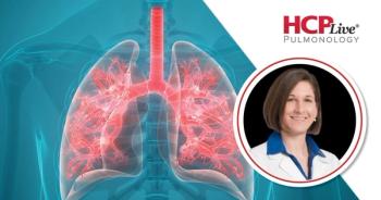
Derek W. Russell, MD
In the hopes of outlining a pathway that contributes to
Study coauthor Derek W. Russell, MD, an assistant professor in the Division of Pulmonary, Allergy, and Critical Care Medicine at the University of Alabama at Birmingham School of Medicine, noted in an interview with MD Magazine® that COPD’s unusual nature comes from the body reacting constantly to harmful inhalants.
“In the US, this usually includes a constant exposure of cigarette smoke, but it can be other irritants [or] pollutants,” Russell said. “The ‘innate’ immune system, cells that are always in our body waiting for bad things to kill, gets overheated and starts to damage the tissues, and in COPD this leads to the spongy lung tissue to die and dissolve, which leaves the lung scarred, floppy, and makes it harder and harder to breathe.”
In cases of COPD, much of the damage appears to be from the innate system, he explained. The body’s own defenses, including proteins that block the dangerous neutrophils’ proteins, can often prevent any harm that results in permanent damage.
Russell and colleagues examined to learn if neutrophil elastase (NE) exists in an exosomal form, and whether these exosomes could potentially bypass alpha1-antitrypsin (A1AT) inhibition to contribute to COPD.
The findings revealed “an unappreciated role for exosomes in the pathogenesis of disorders of extracellular matrix (ECM) homeostasis such as COPD and bronchopulmonary dysplasia (BPD), providing a critical mechanism for proteolytic damage,” authors wrote.
The results of the study concluded that exosomes from activated polymorphonuclear leukocytes (PMN) caused COPD damage when the subcellular particles collected from purified PMNs were injected into the lungs of healthy mice, while exosomes from inactive PMNs did not cause COPD when transferred.
“In the lungs of COPD patients, neutrophils get stimulated by their disease, make NE and it gets coated on the exosomes they are spewing out,” Russell said. “The exosomes with NE on them are much more damaging to lung tissue than the NE that just gets cast to the wind.”
This may be due to the exosomes protecting NE from being blocked by A1AT, as well as because exosomes are able to bind to connective tissue that hold the lung together, he explained. The exosomes are among the major explanations for the “confusing findings” that NE and related enzymes contribute to COPD lung destruction, despite patients’ retained A1AT protective barriers.
The findings of this study have the potential to lead to therapeutic strategies and improved disease monitoring in COPD. Russell added that it seems very likely a similar pathway contributing to other diseases.
“There are many diseases—metastatic cancer, for example—in which the body's own tissues are damaged by enzymes from the body's own cells, but this is not well-understood yet,” Russell said. “We also do not yet know why the neutrophils make these particles.”
He theorized the particles are meant to “seek and destroy” bacteria or tissue, allowing the neutrophils to access an infected part of the body. But any conclusion would require clinical follow-up.
“We are eager for others to build on this research and unravel the effects these tiny particles have in health and disease,” Russell said.
The study, “
Advertisement
Related Content
Advertisement

Latest CME


Advertisement
Advertisement
Trending on HCPLive
1
Q&A: Intensive LDL Lowering and Cardiovascular Prevention Strategies, With Lawrence Leiter, MD
2
Evaluating Clinical and Real-World Evidence in Hypercortisolism
3
Interstitial Lung Abnormalities: Recognition, Risk Stratification, and When to Act
4
Management Strategies for Hypercortisolism
5