Measuring Corneal Nerve Fiber Density May Be Less Invasive Method of Monitoring Peripheral Neuropathy Associated with HIV
Researchers say it may be possible to diagnose and monitor peripheral neuropathy induced by HIV, replacing more invasive methods of testing such as skin biopsy.


Researchers say it may be possible to diagnose and monitor peripheral neuropathy induced by HIV, replacing more invasive methods of testing such as skin biopsy.
Other methods such as electrophysiological testing to measure peripheral nerve conduction properties are not sensitive enough to detect damage to small nerve fibers, especially in early stages of disease.
According to a news release, scientists at Johns Hopkins “developed a simian immunodeficiency virus (SIV)-infected macaque model that closely reflects key peripheral nervous system (PNS) alterations seen in HIV patients with peripheral neuropathy” and used it to determine whether SIV infection leads to corneal nerve fiber loss, and also whether “corneal nerve fiber density correlates with epidermal nerve fiber length counts, thereby setting the stage for follow-up investigation using corneal confocal microscopy.”
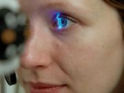
"The cornea is the most densely innervated tissue in the body, so corneal nerve assessment is extremely sensitive for detecting small sensory nerve fiber damage as compared to other tests including measurement of intra-epidermal nerve fibers in the skin,” said Joseph L. Mankowski, DVM, PhD, Professor of Molecular and Comparative Pathobiology, Pathology, and Neurology at the Johns Hopkins University School of Medicine.
The release noted that the methods used by the researchers to measure corneal nerve fiber density “independently demonstrated significantly lower sub-basal corneal nerve fiber density among SIV-infected animals that rapidly progressed to AIDS as compared to slow progressors. Corneal nerve fiber density was also directly correlated with epidermal nerve fiber length.”
“Moving to non-invasive and repeatable methods of nerve fiber measurements such as in vivo corneal confocal microscopy would enhance study of peripheral neuropathy by enabling early detection of damage, progression of nerve fiber deterioration, and enable assessment of therapeutic strategies in the SIV/macaque model," said Mankowski.
He added that “adapting in vivo corneal confocal microscopy for use in tracking HIV-induced PNS damage in patients may be of great value to identify early PNS damage independent of performing skin biopsies.”
Results from the study were discssued in the article “Loss of Corneal Sensory Nerve Fibers in SIV-Infected Macaques,” published in The American Journal of Pathology.


