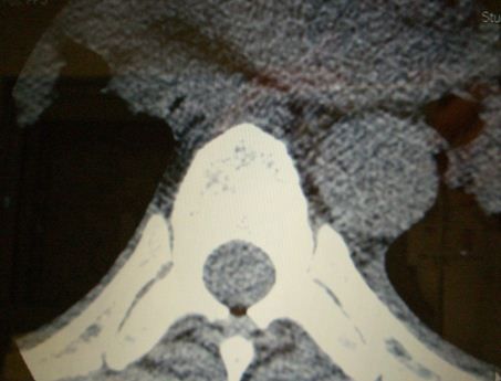
Obese Woman With Fibromyalgia and Worsening Back Pain
This back pain doesn't feel anything like the usual fibromyalgia attack. Neither the chiropractor nor oxycodone helped. Now she's constipated. What diagnosis do you suspect?
An obese woman in her thirties with a history of fibromyalgia syndrome (FMS), polycystic ovarian syndrome, diabetes mellitus (DM) and depression presented to the emergency department with gradually worsening midline back pain of one week's duration. She initially attributed the pain to her FMS, but because it did not improve as usual after a few days, she saw a chiropractor. When this did not help, she visited her physician, who prescribed hydrocodone/acetaminophen and cyclobenzaprine, also to no avail.
For the 24 hours before coming to the emergency department, the patient had been constipated and had difficulty urinating. Both legs began to feel “wobbly” and numb. She described the pain as extending from below her neck down to her waist in the midline, usually worst “just above her bra strap.”
The patient said that this episode definitely was not like her typical FMS attack. She had no other complaints, and when asked specifically, she denied fever, abdominal pain, and vomiting.
On physical examination, the patient’s pulse was 91 beats per minute; blood pressure, 138/76 mm Hg; respirations, 22 breaths per minute, with a pulse oximeter reading of 99%; and temperature, 37.4°C (99.4°F) taken orally.
Findings from inspection of the head and neck were unremarkable, but the astute doctor, suspecting the worst, checked for meningismus, as indicated by the combination of fever and back pain. Chest examination findings were completely normal. Her back and flank areas were not particularly tender. Neither was her abdomen (although she is quite obese).
The neurological examination showed decreased subjective pinprick sensation in both leg, even up to the lower abdomen. The patient had normal distal leg strength with both plantar flexion and dorsiflexion of the foot, but during a straight-leg raise test she could only keep her legs up for one or two seconds.
The radiologist refused to call in the techs for an after-hours MRI scan, opting to begin with a CT scan which he said would “anything of consequence” in the spine. If results were negative, he said, an MRI could always be ordered the next day “if indicated.”

A CT image at the level of greatest pain appearas above. Do you see anything noteworthy? What diagnosis do you suspect?
Click here to see the complete case study.