A Prognostic Test for Knee and Hip Osteoarthritis
A new study indicates that a high bone mineral density test of the femoral neck may serve as an indicator of knee and hip osteoarthritis onset.
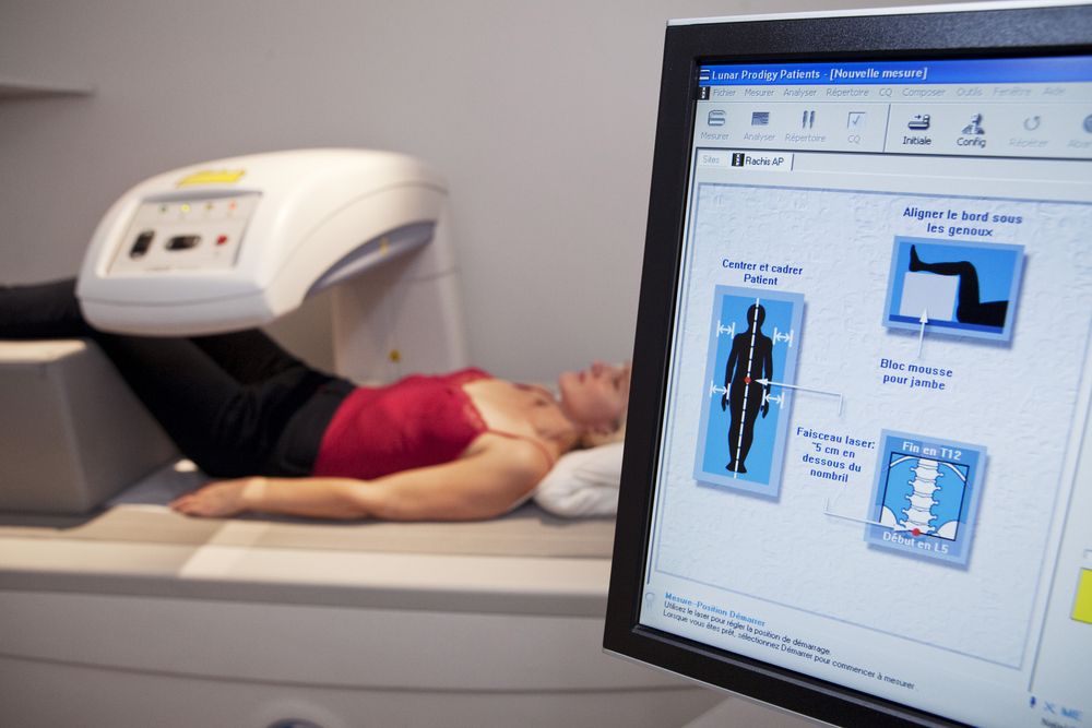
Bone Densitometry Examination (©ImagePointFrShutterstock.com)

A prospective look at the evidence from the Rotterdam Study in elderly men and women further substantiated a previously suggested link between bone mineral density and osteoarthritis.
In a new study by Bergink et al. published in a March issue of Arthritis & Rheumatology, investigators from the original Rotterdam Study added longitudinal support to earlier findings that high bone mineral density of the femoral neck predicts the development of radiographic osteoarthritis of the knee. Additionally, new evidence of a similarly higher associated risk of osteoarthritis of the hip was also reported.
THE STUDY
For the current study, the investigators prospectively evaluated radiographic evidence of the knee, hips, and hands available from patients enrolled in the Rotterdam I study that was originally collected at three visits occurring between 1989-1993, 1997-1999, and 2002-2004, and the Rotterdam II study, with visits between 2000-2001 and 2004-2005. Baseline fracture assessments were also conducted in both populations.
The risk of osteoarthritis of knee was significantly increased by 58 percent in those patients in the highest quartile for bone mineral density compared to those in the lowest quartile. Risk for osteoarthritis of the hip increased by 57 percent in the highest vs lowest quartiles.
The incident risk of osteoarthritis of knee increased by 15 percent for each standard deviation increase in femoral neck bone density, although a similar increase in progression was not observed. The pattern changed with osteoarthritis of the hip, where there was a distinct increase of progression of 16 percent per standard deviation observed.
No increased risk of osteoarthritis of the hand was reported to be associated with increases in bone mineral density or previous history of vertebral fractures. The study did not confirm a protective effect of vertebral fractures on either the incidence or progression of osteoarthritis of the knee.
REFERENCE
Bergink AP, Rivadeneira F, Bierma-Zeinstra SM, et al. Are Bone Mineral Density and Fractures Related to the Incidence and Progression of Radiographic Osteoarthritis of the Knee, Hip, and Hand in Elderly Men and Women? The Rotterdam Study. Arthritis Rheumatol 2019 Mar;71(3):361-369. doi: 10.1002/art.40735. Epub 2019 Feb 9.