Rash Questions for Rheumatologists
Is it a form of lupus, myositis, or a drug reaction? Check your knowledge of skin eruptions and the associated rheumatologic conditions in this brief photo essay.
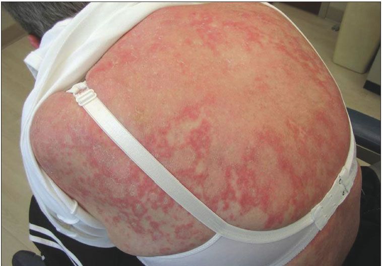
This mildly pruritic rash appeared in late August on the upper back of a 58-year-old woman previously diagnosed with Sjgren syndrome. She had not started any new medications, nor was she taking photo-sensitizing drugs.
The diagnosis: Subacute cutaneous lupus erythematosus.
SCLE is commonly associated with Sjgren syndrome. What is unusual about this woman's presentation?
Where do papules normally appear with SCLE, and what locations are generally spared?
What medications are used to treat it?
For the answers, read: Subacute Cutaneous Lupus Erythematosus
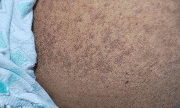
A macular hyperpigmented rash is evident on the abdomen of an 84-year-old African American woman hospitalized with clear vomiting and copious diarrhea of 3-days' duration. She had been diagnosed during a previous hospitalization with multifactorial anemia, stage 4 chronic renal failure, and gouty nephropathy. She had refused nephrectomy, and was discharged on a regimen of 100 mg/d of allopurinol.
At the time of this admission, besides allopurinol, the patient had been taking clopidogrel, amlodipine, metoprolol, acetaminophen, ferrous sulfate, vitamin D3 supplement, pantoprazole, and hydralazine.
The rash is an indication of allopurinol sensitivity, also known as A-DRESS (allopurinol drug-induced rash with eosinophilia and systemic symptoms).
What is most noteworthy about A-DRESS as compared to the other DRESS syndromes?
What other factors in this patient's history contributed to the development of A-DRESS syndrome?
What is the normal course of the rash after allopurinol is discontinued?
Find the answers to the above questions in one of two case reports about the syndrome in this article:
Understanding Allopurinol-Induced DRESS Syndrome
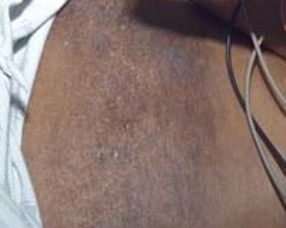
This image shows the the "shawl sign" characteristic of dermatomyositis: a diffuse, flat, erythematous lesion that occurs over the upper chest and back.
What are the defining pathological differences between dermatomyositis and polymyositis?
What should be the chief concern in treating dermatomyositis, and what medications would you prescribe?
For the answers, see:
Diffuse Macular Hyperpigmented Rash and Weakness in an African American Woman
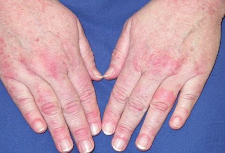
These hands display Gottron papules, pink or vilaceous papular lesions on the dorsal aspects of the distal interphalangeal joints. Also sometimes seen on the elbows, knees, and ankles, these eruptions are also typical of dermatomyositis, and may precede muscle weakness.
This review focuses on the many inflammatory muscle diseases that are associated with weakness: Diagnosis and Management of Inflammatory Muscle Disease
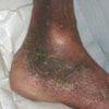
The 68-year-old man who had this rash presented with a two-month history of progressive weakness, arthralgia, and myalgia, and this developing rash. Close examination showed petechial lesions and follicular hyperkeratosis with perifollicular hemorrhage and corkscrew hairs.
The patient also had poor dentition and swollen, purple, spongy gingivae.
The diagnosis was scurvy. Although unusual in developed countries, this diagnosis may arise occasionally. This patient revealed that his diet consisted entirely of potatoes, because he lived alone and did not know how to cook.
Very likely you can guess the treatment and the resolution of this case, but it may be worthwhile taking a close look at the images in this brief case study: Scurvy

The "irritation" at left had persisted for several months in this 46-year-old man's groin. Close observation revealed a tender, macerated, hypopigmented plaque at the junction of the scrotum and upper inner thigh, with some erythema at the periphery of the lesion and several small, superficial erosions within the plaque.
This patient has discoid lupus erythematosus. While the disorder rarely presents in this location, its manifestations can occasionally arise where the sun doesn't shine.
What are the treatment options?
Is it important for this patient to avoid sun exposure?
For the answers, refer to: Discoid Lupus Erythematosus
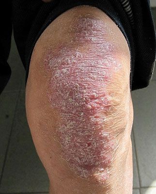
The psoriasis on this patient's knees had persisted for two decades.
He presented at the age of 44, as a recent immigrant from Taiwan.
This rash was associated with a comorbid condition that eventually proved fatal.
From the bare facts above, can you guess the nature of the disease that ultimately led to his demise?
For the answer, see: Skin Eruption in a Taiwanese Immigrant