Vascular Calcification Shown on X-Ray Assesses Cardiovascular Risks in Chronic Kidney Disease
A recent study presented at Kidney Week 2013 suggests that a plain X-ray is a reliable and inexpensive tool for assessing the risks of cardiovascular events and death in patients who have chronic kidney disease (CKD) and are not on dialysis.


A recent study presented at Kidney Week 2013 suggests that a plain X-ray is a reliable and inexpensive tool for assessing the risks of cardiovascular events and death in patients who have chronic kidney disease (CKD) and are not on dialysis.
At the American Society of Nephrology's annual meeting, held November 5-10, 2013 in Atlanta, GA, Pablo Molina, MD, and colleagues from the Department of Nephrology of the Hospital Universitario Dr. Peset in Spain explained that vascular calcification (VC) is extremely prevalent in CKD patients who are not dialyzed. Although it’s well known that the presence of VC is associated with poor prognosis in patients on dialysis, the impact of VC during earlier stages of CKD is not well understood. In fact, only 1 study in the medical literature has described the long-term impact of coronary calcification on CKD patient survival, and no other data is available regarding the effect of VC in other vascular areas.
The goals of Molina’s OSERCE-II study were to evaluate the prevalence of VCs seen on plain X-ray of the hands, hip, and abdomen in non-dialysis CKD patients; correlate VC with other known cardiovascular risk factors; and learn the value of VC as a predictor of future hospitalization, renal and cardiovascular events, and death.
The recent multicenter, observational, prospective OSERCE-II study enrolled 722 non-dialysis CKD subjects from 39 nephrology clinics throughout Spain between 2009 and 2012. The mean patient age was 66±13 years, and 35% of patients were women, while 37% of patients had diabetes mellitus. Overall, 40% of patients had stage 3 CKD, 46% had stage 4, and 14% had stage 5.
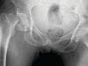
VCs were assessed at baseline according to Adragao scores (AS) obtained from X-rays of the pelvis and hands, as well as Kauppila scores (KS) obtained from X-rays of the lateral lumbar spine. Blood pressure, Ankle-brachial index, and biochemical values were measured, and cardiovascular and renal events and hospitalizations were recorded.
The researchers examined approximately 2,200 X-rays 2 times each. For validation, a subset of films was read later for inter-rater agreement (kappa: 87.3), concordance in groups (92%), and concordance in final score (83%).
VCs were found in 79% of the patients, and they were prominent in 47% (AS ≥3:30%; KS >6:31%). During a 3-year observational period, 74 (10%) deaths occurred, 174 (24%) patients required hospitalization and 154 (21%) patients began dialysis treatment. The most common cause of death was cardiovascular (42%).
According to multivariate analysis, the factors independently associated with mortality were age (odds ratio [OR] 1.067; P <0.001), diabetes (OR 1.738; P =0.049) and AS ≥3 (OR 2.130; P =0.008). Independent predictors of hospitalization were diabetes (OR 1.880; P <0.001], low albumin levels (OR 0.661; P =0.005) and KS >6 (OR 1.524; P =0.024). Patients with VC were not at increased risk for requiring dialysis, the study authors said.
The researchers concluded that VC is highly prevalent among CKD patients who are not undergoing dialysis, and standard X-ray, which is reliable and inexpensive, can help detect the risk of cardiovascular events and death in CKD patients.
The authors disclosed that Abbvie sponsored their study.


