3-D Printed Heart Models on the Rise
The study of complex congenital cardiac anatomy has previously been limited to 2-dimensional assessment. Now the potential use of 3-dimensional cardiac models could allow for previously unavailable visualization and analysis of anatomy.


While the study of complex congenital cardiac anatomy had long been limited to 2-dimensional (2-D) assessment, the potential use of 3-dimensional (3-D) cardiac models could reveal previously unavailable visualization and analysis of anatomy.
New research presented at the 2014 American Heart Association (AHA) scientific Sessions reported an experimental 3-D printed heart model using plaster or ceramic to potentially help surgeons treat patients born with complicated heart disorders.
Matthew Bramlet, MD, the study’s lead author, assistant professor of pediatric cardiology, and director of the Congenital Heart Disease MRI Program at the University of Illinois College of Medicine, shared, “With 3-D printing, surgeons can make better decisions before they go into the operating room. The more prepared they are, the better decisions they make, and the fewer surprises that they encounter.”
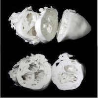
Actual heart models provide a new dimension of understanding beyond the scope of 2-D or even 3-D images.
Researchers chose 3 patients, a 9-month-old girl, 3-year-old boy, and woman in her 20s, all with complex spatial orientation surgical problems. Inexpensive plaster composite material was utilized to create heart models that consisted of 3 components in a short axis orientation, a basal portion, a middle slice containing all four valves, and an apical portion. After studying the models and traditional images, surgeons successfully restored all 3 patients’ severe heart abnormalities.
“You could see that if you make this compromise here, you could fix this problem, and go from a single-ventricle to a 2-ventricle pair. That is the difference, potentially, between a life expectancy of 2 to 3 decades, to 4, 5, or 6, decades,” said Bramlet.
The study concluded low cost, solid opaque cardiac models with intricate intracardiac details improve surgical planning in complicated CHD.
Experts warned 3-D printing was a small study, therefore an emerging technology not yet approved by the Food and Drug Administration.


