
Opinion|Videos|August 31, 2023
Approaches to Imaging in oHCM
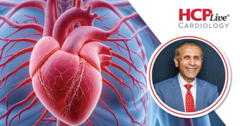
Milind Desai, MD, MBA, leads a discussion on the importance of accurate imaging in oHCM and aortic stenosis, highlighting the echocardiogram and doppler tests.
Advertisement
Episodes in this series

Anjali Owens, MD: Let me shift gears a little bit, but talk a little bit more about the imaging and the importance of accurate imaging. You mentioned the dynamic outflow tract obstruction that's present in HCM [hypertrophic cardiomyopathy] as opposed to the more fixed obstruction is present in aortic stenosis and that some patients may have both or you know, 2 levels of obstruction. What is your approach to imaging in terms of understanding those patients, differentiating them and then treat?
Milind Desai, MD, MBA: So it is important, absolutely important to have a precision in understanding of that area. The LV outflow tract [LVOT] and the aortic valve disease. I would say echocardiography is the mainstay, both the 2D-echocardiography as well as Color Doppler as well as spectral Doppler. The combination of these things do is help us understand what's happening at the LVOT level. Let's say for example the spectral Doppler you can use continuous wave or pulse wave Doppler to define a hub that the obstruction is in the line of the outflow tract. And then the spectral Doppler will help you decide where the obstruction is. Is it in the outflow tract? Is it at the aortic valve? Is it somewhere else in the mid cavitary region? So and you know anytime I'm looking at these scenarios so here are the things I'm looking for. Can I see the valve? If not, we need to work hard to see the aortic valve. Are the leaflets opening? Is there calcification except? If the valve is opening. What's happening in the outflow tract? Is the outflow tract narrow? Are you seeing a subaortic membrane? The other obviously more common thing is what's happening with the mitral valve? Are you noticing something systolic anterior motion of the mitral valve where it is making septal contact during systole, which is the hallmark of hypertrophic obstructive cardiomyopathy. If it is doing so, then what? Is the corollary Doppler and how high is the gradient? Anything more than 30 at rest is significant. The other important aspect of HCM is this is` dynamic. So you can use different maneuvers—Valsalva, stress echocardiography, amyl nitrite inhalation in many labs to provoke the gradient. It is a provokable gradient that is commonly seen in in HCM. The other important thing not just to stop at whether or not there's provokable gradient, but what is causing it. Is it driven mostly by the septal wall thick? Is it, mitral valve adding fuel to the fire. Is it papillary muscles adding to the fuel, fuel to the fire? So you have to understand that the dynamic interplay of everything, one last thing I'm going to say is basically that of subaortic membrane that so aortic stenosis has a continuous-wave Doppler profile fixed obstruction. HCM has a late peaking dynamic profile. Subaortic membrane tends to behave a lot like aortic stenosis, and so if you have a normally functioning valve but a fixed Doppler profile, that's you have to look for subaortic membrane. So you have to you have to go through progressions and work your differential diagnosis.
Anjali Owens, MD: I think another thing not to forget that's in our toolbox is an invasive hemodynamic assessment. So Andrew, being an interventional cardiologist, what do you see the role as in terms of differentiating AS and dynamic outflow tract obstruction in the cardiac catheterization laboratory?
Andrew Wang, MD: Yeah, it's challenging because it is hard to localize what is the site that you're measuring in the left ventricle with a catheter where there are certain catheters that we can use to try to identify what is truly intracavitary versus a valvar gradient, but it can still be very challenging. I actually think that the noninvasive imaging is as good as we have to differentiate and as you mentioned, sometimes it's a combination of both in which that case, treatment of 1 may not improve the patients symptoms to the degree that you want and so you really have to think of it as a continuous or combination condition.
Anjali Owens, MD: There are a handful of patients a year where we end up doing a concomitant septal myectomy and aortic valve replacement. You know, certainly not the most common, but a handful year.
Milind Desai, MD, MBA: A few years ago, we published our data on almost 200 such patients who underwent a VR and a myectomy. The outcomes were pretty similar to an A gender mass population in at least in an experience center. So it is important poor form to leave a disease diuretic valve on the table, or a or A or vice versa.
James Januzzi, MD: Vice versa, yeah.
Milind Desai, MD, MBA: That happens more often than not.
James Januzzi, MD: In fact, for the viewers, I think it's important to recognize relief of LV outflow tract obstruction in the form of an AVR could theoretically be fraught with production of an LVOT gradient because you no longer have afterload and so post operative catastrophes of patients with acute systolic anterior motion of the mitral valve in people with unrecognized HCM at the time of an AVR have been reported. So really carefully assessing the culprits in LVOT obstruction in these patients is really I think crucial and the management is driven postoperatively to some extent as well, because a patient like that, you're not going to put it on an inotrope if they're in shock, they need afterload. So it really is a an uncommon but potentially high-risk complication.
Milind Desai, MD, MBA: Andone tidbit I'm going to mention about this. Not necessarily HCM, but if you have patience with your severe chronic aortic insufficiency with a big, dilated LV and they have developed some element of hypertrophy as a way of compensation, you fix their aortic valve, replace it, the heart shrinks. Four months later, they come back short of breath because now they have a hypertrophic heart and they're starting to send. That is, I would say behaves like HCM but not really. We've had in my career I've had a few such patients referred to us or it's not a failed prior procedure it's just a ramp downstream ramification of something like that happened.
Anjali Owens, MD: It just speaks to the importance of following these patients longitudinally and really understanding the underlying, you know, pathophysiology.
Transcript is AI-generated and edited for clarity and readability.
Advertisement
Related Content
Advertisement


Latest CME


Advertisement
Advertisement
Trending on HCPLive
1
Prevalence and Recognition of Hypercortisolism
2
Screening and Diagnosis of Hypercortisolism in Type 2 Diabetes
3
Distinguishing CSU From Chronic Inducible Urticaria Subtypes
4
Diagnosing the Hallmark Signs and Symptoms of CSU
5