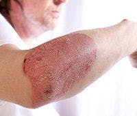
Fine Line Between Clear and Almost Clear Psoriasis Severity
Understanding the difference between clear and almost clear skin is crucial with the advent of new biologic therapies that make complete clearance attainable.

Understanding the difference between clear and almost clear skin is crucial with the advent of new biologic therapies make complete clearance attainable.
Researchers presented comparisons in patient perceptions of the severity of psoriasis signs of moderate-to-severe plaque psoriasis that achieved complete skin clearance (sPGA 0) and almost clear skin (sPGA 1) based on Psoriasis Symptom Inventory (PSI) scores at the American Academy of Dermatology 2015 Annual meeting in San Francisco, CA.
Physicians routinely assess psoriasis severity with the static Physician’s Global Assessment, through which treatment response is typically defined as achieving a sPGA of 0 or 1.
A total of 295 adult patients with moderate-to-severe psoriasis receiving a biologic were enrolled in this cross-sectional, observational study. Upon completing 8-item PSI electronic daily diary on 7 consecutive days (Day 1-7), changes in sPGA status in some patients excluded 65 individuals.
Of the sPGA 0 subjects, 60.8% reported a PSI=0 compared with 5.3% who had sPGA 1. Furthermore, approximately 95% of sPGA 0 patients met the PSI responder threshold compared with 54.3% with sPGA 1.
Researchers concluded, “A significantly higher proportion of patients with skin clearance (sPGA0) reported no psoriasis symptom severity compared to patients rated as ‘almost clear’ (sPGA 1). A significantly higher proportion of sPGA 0 patients (95%) also achieved not at all (0) or mild (1) psoriasis severity on all 8 PSI signs and symptoms compared with sPGA 1 patients (54%).”
Physician assessments indicated, patients who were considered ‘clear’ reported either no severity or significantly lower severity of psoriasis signs and symptoms than those rated as ‘almost clear’.


