Impact of Progression of Diastolic Dysfunction on Mortality in Patients With Normal Ejection Fraction


O. Julian Booker, MD
Review
Aljaroudi W, Alraies MC, Halley C, et al. Impact of progression of diastolic dysfunction on mortality in patients with normal ejection fraction. Circulation 012;125(6):782-788.

doi:10.1161/CIRCULATIONAHA.111.066423
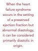
Diastolic dysfunction is the abnormal myocardial mechanical process that occurs during diastole. Heart failure is the clinical syndrome characterized by increased total body fluids and decreased ability to provide adequate tissue perfusion. Diastolic dysfunction can occur in conjunction with or in isolation from systolic dysfunction as measured by ejection fraction. When the heart failure syndrome occurs in the setting of a preserved ejection fraction but abnormal diastology, it can be considered primarily diastolic in origin.
Study Details
Consecutive patients from a single center found to have a left ventricular ejection fraction (LVEF) ≥55% by echocardiography from January 1, 2005, through December 31, 2009, were reviewed. Patients having a second echocardiogram within 6 to 24 months were selected for analysis. Patients with prior mitral valve surgery, severe mitral stenosis, or regurgitation were excluded. Those conditions that confound the assessment of diastology, such as atrial fibrillation, tachycardia, and incomplete examinations, were also grounds for exclusion, but not all patients with atrial fibrillation were excluded. Those persons without a valid social security number were
not included in the analysis. Of the 3223 patients with 2 echocardiograms within the allotted period, 1065 were selected for inclusion.
Echocardiography was performed on commercially available systems across multiple vendors. Diastolic function was determined using a combination of mitral inflow pattern, annular tissue Doppler velocities, and pulmonary vein flow pattern in accordance with the recommendations by the Doppler Quantification Task Force of the Nomenclature and Standards Committee of the American Society of Echocardiography.1 In those cases where diastology was assessed in the setting of atrial fibrillation, a combination of mitral inflow deceleration time and tissue Doppler parameters was used. Diastolic dysfunction was then graded as mild (grade 1, impaired relaxation), moderate (grade 2, pseudonormal), or severe (grade 3, restrictive). Systematic review of echocardiograms was not performed, and all data were taken from prior reads.

All clinical data were acquired from review of the electronic medical records system. Ethnically, the patient population was fairly representative of the United States population, and the breakdown of cardiac risk factors was also highly representative. The most common indications for testing were symptoms, valve disease, and ventricular function, and were typical of what we usually see in clinical practice. The patients’ medical regimens at baseline and at the time of the subsequent echocardiogram were similar. As seen in other studies, there was an association between diastolic dysfunction and both age and hypertension.
Two-hundred ninety patients were thought to have normal diastolic function, with 88% of these being classified as having an essentially normal echocardiogram. Some degree of diastolic dysfunction was seen in 72% of patients, with mild disease being the most common, comprising 65% of the subjects Only 7.4% of the included patients were felt to have moderate or severe disease. At the time of the follow-up exam, 73% had stable disease, whereas 16% had worsening and 11% had improvement of diastolic function. Of note, 88 patients (8.3%) had a decrease in LVEF to <55%.
All-cause mortality was determined by way of the social security death index with a censuring date of June 1, 2011. A total of 142 patients died during a period of 1.6 ± 0.8 years following their second echocardiogram. Of those patients, 20 had normal diastolic function, 102 had mild dysfunction, and the final 20 had what was considered
moderate or severe dysfunction. Any worsening in diastolic function either from normal to abnormal or from a previously abnormal result to a higher grade of dysfunction portended a worse prognosis compared with those persons having no change or improvement. Additionally, any fall in the LVEF into the abnormal range negatively affected mortality. On the whole, there was no difference in mortality between those persons with improved diastolic function and those without change. However, patients
with improvement of at least 2 classes showed a statistically insignificant survival benefit compared with those having more modest improvement.
References
1. Quinones MA, Otto CM, Stoddard M, Waggoner A, Zoghbi WA. Recommendations for quantification of Doppler echocardiography: a report from the Doppler Quantification Task Force of the Nomenclature and Standards Committee of the American Society of Echocardiography. J Am Soc Echocardiogr. 2002;15:167-184.
2. Dokainish H, Zoghbi WA, Lakkis NM, et al. Optimal noninvasive assessment of left ventricular filling pressures: a comparison of tissue Doppler echocardiography
and B-type natriuretic peptide in patients with pulmonary artery catheters. Circulation. 2004;109:2432-2439.
3. Halley CM, Houghtaling PL, Khalil MK, Thomas JD, Jaber WA. Mortality rate in patients with diastolic dysfunction and normal systolic function. Arch Intern Med.
2011;171:1082-1087.
4. Okura Y, Ohno Y, Ramadan MM, Suzuki K, et al. Characterization of outpatients with isolated diastolic dysfunction and evaluation of the burden in a Japanese community: Sado Heart Failure Study. Circ J. 2007;71:1013-1021.
5. Tehrani F, Morrissey R, Phan A, Chien C, Schwarz ER. Statin therapy in patients with diastolic heart failure. Clin Cardiol. 2010;33:E1-5.
6. Jassal DS, Bhagirath KM, Karlstedt E, et al. Evaluating the effectiveness of rosuvastatin in preventing the progression of diastolic dysfunction in aortic stenosis: a
substudy of the aortic stenosis progression observation measuring effects of rosuvastatin (ASTRONOMER) study. Cardiovasc Ultrasound. 2011;7;9:5-12.
7. Chang SA, Kim YJ, Lee HW, et al. Effect of rosuvastatin on cardiac remodeling, function, and progression to heart failure in hypertensive heart with established left
ventricular hypertrophy. Hypertension. 2009;54:591-597.
8. Okura H, Asawa K, Kubo T, et al. Impact of statin therapy on systemic inflammation, left ventricular systolic and diastolic function and prognosis in low risk ischemic
heart disease patients without history of congestive heart failure. Intern Med (Tokyo, Japan). 2007;46:1337-1343.
Commentary
Moderate or Severe Diastolic Dysfunction Calls for Aggressive Risk-Factor Modification
In this retrospective study, a standardized echocardiographic assessment of diastolic function was performed in patients with normal ejection fractions. The authors’ interpretation of diastolic function relied upon real-world indices that are routinely acquired during any clinical echocardiogram and were composed of primarily mitral
inflow pattern, annular tissue Doppler velocities, and pulmonary vein flow pattern.

Diastolic dysfunction, with or without heart failure, can be assessed with echocardiography. Dokainish et al2 showed rather elegantly that in the ICU setting, E/Ea (E/e'), a measure of global left ventricular diastolic function, correlates well with pulmonary capillary wedge pressure in the subgroup with preserved LVEF. It has also been shown that higher levels of isolated diastolic dysfunction are associated with a higher mortality (Halley et al),3 irrespective of the presence or absence of heart failure symptoms (Okura et al).4 The current study took another step forward and determined that any degree of diastolic dysfunction confers a higher degree of mortality. Equally important, a marked improvement in diastolic function may improve overall prognosis. Data show that patients with heart failure with preserved ejection fraction benefit from medical therapy. The correlation between abnormal diastology and left ventricular end-diastolic pressures has been well documented. Taken together, it becomes clearerthat moderate or severe diastolic dysfunction should be treated by aggressive risk-factor modification. The increase in mortality associated with all levels of diastolic
dysfunction shown in the current study extends the use of medical therapy organically toward the treatment of even mild dysfunction, specifically the use of statins.5-8

Some authors have expressed concerns related to Doppler echocardiography’s ability to accurately measure left ventricular diastolic function. Catheter-based measures of diastolic function, such as the time constant of LV relaxation, remain the gold-standard methods to assess global myocardial function, but are invasive. Arguments can be made that other measures of myocardial relaxation may provide a more accurate, less load-dependent measure of diastology than does the standard Doppler evaluation. Tagging with cardiac MRI and speckle tracking by echocardiography are able to quantify myocardial deformation using metrics such as strain or strain rate, in a segmentby- segment basis, but these measures have not proved to be more powerful predictors of clinical outcomes. When compared with some of the aforementioned methods, the measures utilized in the current study might not be as sensitive to smaller degrees of diastolic dysfunction, but have proved themselves to be perhaps more clinically useful and powerful prognostic tools.
The authors mention several limitations of their study: (a) the study is retrospective and from a single institution, (b) readers were not blinded to medical history of patients, (c) there was difficulty in distinguishing normal from pseudo-normal mitral inflow patterns, (d) left atrial volumes were unavailable in many patients, (e) tissue Doppler imaging parameters were not available in some patients (exact percentage not given), (f) some patients with atrial fibrillation, in whom it is difficult to reliably assess diastolic dysfunction, were included in the study, (g) there were a relatively small number of patients with moderate dysfunction, and only 2 with severe diastolic dysfunction, and (h) cardiac death or hospitalization was not assessed in the outcome data. In addition, the authors mention that LVEFs were evaluated quantitatively “and/or” visually, but the number of patients evaluated only visually is not provided. In these patients with visually estimated LVEF, the delineation of preserved LVEF may be an issue. It also precludes the evaluation of LV volumes in these patients.
Lastly, the investigators used a motley combination of mitral inflow, tissue Doppler, and pulmonary vein parameters to classify diastolic dysfunction, but the relative weight assigned to each parameter is not provided. The absence of having pre-set, well-defined parameters also complicates evaluation of interobserver agreement, which
was only 83% in this study. Other parameters that would confound detection of LV diastolic dysfunction—such as heavy mitral annular calcification resulting in high mitral early diastolic velocities— were not taken into account (most of their patients were elderly). Using mitral inflow velocities to detect grade 1 diastolic dysfunction has limited reliability, as it is dependent on myriad technical and other factors including heart rate and loading conditions. To what extent these limitations impact or decrease the validity of the study’s findings is difficult to determine, but the fact that the investigators analyzed and followed over 1000 patients lends a reasonable measure of credibility
to their conclusions.
About the Authors
O. Julian Booker, MD, is assistant professor of medicine in the Division of Cardiovascular Disease at the University of Alabama at Birmingham. Dr Booker’s medical degree is from Baylor College of Medicine, Houston, TX, where he also completed his internship, residency in internal medicine, and a fellowship in cardiovascular disease. He completed fellowships in cardiac MRI and CT at the National Institutes of Health. Dr Booker was assisted in writing this article by Navin C. Nanda, MD, Distinguished Professor of Medicine and Cardiovascular Disease and director of the Heart Station/Echocardiography Laboratories of the University of Alabama at Birmingham. Dr Nanda is also director of the Echocardiography Laboratory at the Kirklin Clinic, University of Alabama Health Services Foundation. He has received many awards for service and achievement in medicine and is the author of numerous publications.