OA and RA Treatment: Targeting MGC Cells May Be a Promising Avenue
A study in BMC Musculoskeletal Disorders suggests that multinucleated giant cells (MGC) may contribute to osteoarthritis (OA) and rheumatoid arthritis (RA) in addition to their known association with synovitis severity. The finding adds to other recent research and points to the therapeutic potential of targeting MGCs to improve pain and joint damage in both types of arthritis.


A study in BMC Musculoskeletal Disorders suggests that multinucleated giant cells (MGC) may contribute to osteoarthritis (OA) and rheumatoid arthritis (RA) in addition to their known association with synovitis severity. The finding adds to other recent research and points to the therapeutic potential of targeting MGCs to improve pain and joint damage in both types of arthritis.
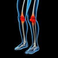
MGCs have been described in diverse arthritic conditions for some time, but their role in the pathogenesis of OA and RA remains unknown. Changes in the synovium and subchondral bone are closely associated with symptoms of OA and are thought to contribute to cartilage damage; synovitis predicts structural severity and progression of tibiofemoral cartilage damage in OA. Synovitis in RA drives pain, cartilage degradation and pannus formation with subsequent erosions. Subchondral inflammation might also contribute to increased bone turnover and joint damage in both OA and RA.
“Although OA and RA differ in their etiology, with mechanical factors being key to knee OA progression and specific immunity driving RA, common mechanisms may contribute to joint damage and pain in both conditions,” the study authors noted.
The study measured knee synovial and subchondral bone samples from age-matched patients selected from the Arthritis Research UK Pain Centre Joint Tissue Repository who underwent total joint replacement for OA or RA, versus non-arthritic post mortem (PM) controls. OA synovium was stratified by histological inflammation grade using index tissue sections. Synovitis was assessed by Krenn score. Synovial MGCs were identified as large cells showing three or more nuclei inside their cytoplasm in haematoxylin and eosin stained sections, thereby avoiding confounding by mitotic cells with two nuclei. Inflamed OA and RA synovia displayed more multinucleated giant cells than did non-inflamed OA and PM synovia. There was a significant association between MGC numbers and synovitis severity.
“Together, our findings indicate a clear activation of mononuclear phagocyte system cells, both in synovium and in subchondral bone, and both in OA and in RA,” the authors revealed. “Synovial inflammation is a common feature of OA, although the intensity and nature of inflammation may differ between OA and RA. It is currently unknown whether synovitis characterizes a subgroup of people with OA, or may occur during phases of the disease in all patients. In cross sectional studies, the extent of synovitis varies between cases, ranging from normal to severe inflammation.”
In addition, the study found that there is a prevalent subgroup of people with OA in whom the numbers of MGCs are comparable to those in people with RA at the time of joint replacement surgery. Furthermore, MGC densities were significantly associated with the total synovitis score indicating that this is a cellular characteristic of synovitis that is shared between OA and RA, suggesting shared inflammatory mechanisms.
Further research will be needed to establish whether synovial inflammation and subchondral bone remodeling each may be regulated by different mediators.