OCT Angiography Provides Vivid Pictures of Nonproliferative Diabetic Retinopathy Severity
Speaking at the American Academy of Ophthalmology 2016 Meeting in Chicago, K. V. Chalam, MD, expressed the advantage of using non-invasive OCT angiography (OCT-A) as opposed to the older fluorescein angiography as essentially a difference between imaging in three dimensions or two.

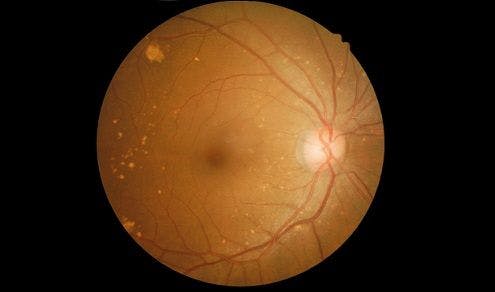
Speaking at the American Academy of Ophthalmology 2016 Meeting in Chicago, K. V. Chalam, MD, expressed the advantage of using non-invasive OCT angiography (OCT-A) as opposed to the older fluorescein angiography as essentially a difference between imaging in three dimensions or two. In application, he was discussing how accurately Split Spectrum Amplitude Decorrelation Angiography (SSADA)-assisted OCT-A could detect nonproliferative diabetic retinopathy (NDPR).
The study looked at 60 normal eyes, 36 with mild NDPR, 21 with moderate NDPR, 13 with severe NDPR, and 32 eyes that had both NDPR and diabetic macular edema (DME). Excluded from the study were those with glaucoma or cataract. The imaging allowed them to observe deep perifoveal vessel density with a great degree of detail. All imaging was administered by a single, experienced OCT-A operator.
When deep capillary macular vascular flow was observed and graphed, results displayed a very sharp linear decline as the seriousness of NDPR increased, with a six fold drop in density from normal eye to severe NDPR.
The study showed the specificity and effectiveness with which OCT-A can express the seriousness of NDPR. Towards the conclusion of his presentation, Chalam suggested that the technology may be of clinical use in assessing the severity of NDPR that would otherwise go unnoticed, or perhaps as a way of tracking the progress of treatments.
Related Coverage
Assessing Rates of Noninfectious Vitritis after Intravitreal Injection of Anti-VEGF Agents
Amsler vs. Casa: Which Test Better Detects Early Wet AMD?
Difluprednate Shows Itself Effective for Intermediate Uveitis Treatment


