Patients with Parkinson's Disease Have Worse Eyesight than Healthy Patients
Parkinson's disease patients (PD) have significantly worse vision for low contrast images at close and far distances, according to findings published in the Journal of Parkinson's Disease. At far distances, the PD patients had deficient abilities viewing high contrast images, too.


Parkinson’s disease patients (PD) have significantly worse vision for low contrast images at close and far distances, according to findings published in the Journal of Parkinson’s Disease. At far distances, the PD patients had deficient abilities viewing high contrast images, too.
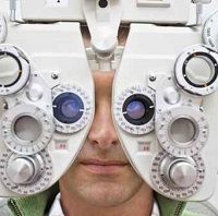
An international team of researchers observed 32 patients with PD and 71 control subjects to evaluate low and high contrast letter acuity using a variable contrast acuity eye chart developed for the Apple iPad. The subjects viewed the Variable Contrast Acuity Chart on an iPad with both eyes at a close distance (40 cm) and a far distance (2 m). The chart was also viewed at high contrast (black and white visual acuity) and 2.5 percent low contrast (grey on white). The patients additionally received a neurological examination, including the Unified Parkinson’s Disease Rating Scale (UPDRS). If the patients required corrective lenses, they were permitted to use them for the study.
The researchers found that after analysis with a visual acuity scale based on the number of letters the study subjects correctly identified, PD patients could see about 10 percent fewer letters than the non-PD control subjects in the low contrast tests at both the close and far distances. In the high contrast setting, this difference was observed only in the far distance test. The researchers wrote there was no statistical difference between the PD and non-PD test subjects in the high contrast test at 40 cm.
“Visual impairments can have a significant impact on quality of life and day to day functions,” lead investigator Charles H. Adler, MD, PhD, Department of Neurology, Mayo Clinic College of Medicine, Scottsdale, AZ, explained in a press release. “However, impaired contrast sensitivity in PD patients is a topic that has received relatively little attention in clinical practice. The electronic form of low contrast acuity letter chart we used in this study combines contrast sensitivity with visual acuity testing and is quick and easy to administer. It is portable, quantitative, and adjustable for testing distances and contrast levels.”
The authors added that if the iPad were to be implemented as a screening tool, it would lead to better visual testing for PD patients.
“We believe that an assessment of low contrast acuity should be considered as part of the standard eye exam in PD, as it may reveal an otherwise missed visual impairment in these patients, help clinicians define a set of criteria for counseling them regarding safety while walking and driving at low light conditions, and guide both pharmacologic and non-pharmacologic interventions that increase functional capacity,” Adler concluded.


