Article
Scleroderma Breakthrough Offers Novel Therapeutic Target
Author(s):
Investigators from Milan, Italy, have identified an important molecule involved in the onset of systemic scleroderma.

An important molecule involved in the onset of systemic sclerosis, or scleroderma, has been identified, according to a new report. The rare autoimmune disease—which causes inflammation, occlusion of the small blood vessels, and fibrosis in the skin and internal connective tissues—does not have any known effective treatments. The fibrosis could eventually lead to severe complications for the patient, but the study authors believe that their work might offer hope for a novel therapeutic target.
Investigators from Milan, Italy, investigated the mechanisms that could contribute to endothelial damage in patients in order to understand the pathology of scleroderma. The study authors hypothesized that platelets might be a source of high-mobility group box 1 (HMGB1) protein in scleroderma patients, which they thought could be critical to sustaining and promoting vascular damage.
The platelet HMGB1 protein activates neutrophils and encourages them to create neutrophil extracellular traps (NETs), the investigators explained. The NETs are a source of autoantigens and are relevant in autoimmune diseases; however, little is known about their effects on scleroderma patients. The investigators suspected NETs were involved and, as such, set out to verify their function.
For their study, investigators collected blood samples from 57 scleroderma patients and 59 control subjects to analyze their expression of HMGB1, among other data. They also studied the NET by-products in the blood plasma.
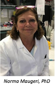
“We showed both in vitro and in an animal model of the disease that the presence of microparticles expressing HMGB1 is enough to activate the immune system and in particular neutrophils,” the study’s first author, Norma Maugeri, PhD, said in a press release she provided to Rare Disease Report®.
The investigators observed that the scleroderma patients expressed the platelet-specific CD61 antigen in both the whole blood and platelet free plasma compared with healthy volunteers. The scleroderma patients were found to be more likely to express damage-associated molecular patterns of HMGB1 than the healthy patients included in the study. The investigators also noted that activated autophagic neutrophils were “abundant” in the blood of scleroderma patients, where they were not in healthy patients. Thus, they extrapolated that they are involved in endothelial damage and fibrosis in scleroderma patients.

When these microparticles derived from scleroderma patients were injected into mice, neutrophil and NET production were activated and caused endothelial damage. However, microparticles from healthy control subjects did not cause endothelial damage after having been injected into the mice models.
“Endothelial damage is an early and critical feature of scleroderma, and our findings suggest that platelet activation is an upstream factor that contributes to the pathogenesis of scleroderma,” the study authors write.” The cause of platelet activation in scleroderma patients, though, remains to be determined.”
While the initial trigger has yet to be identified for scleroderma patients, the study authors write that the “vicious circle of platelet activation is maintained” through endothelial damage exposing collagen from the subendothelial matrix. Possible scleroderma biomarker candidates in the future include platelet microparticles, activated and autophagic neutrophils and NET byproducts, as those for scleroderma and systemic lupus erythematosus cannot yet be separated.
Once other studies are able to replicate these findings, the investigators said HMGB1 may become the first therapeutic target for scleroderma. One way to do this would be to interrupt the platelets that release HMGB1 or eliminating it entirely to slow the onset of the disorder.
“Obviously the results we obtained must be further confirmed and expanded, yet we believe that an excessive amount of HMGB1 outside the cells might be the main cause of damage to vessels and fibrosis of connective tissues,” study author Angelo Manfredi, MD, concluded in his statement.
Feature Picture Caption: Platelet derived microparticles (red) expressing HMGB1 activate neutrophils, inducing autophagy (green), citrullinization of DNA (blue) and the release of neutrophil extracellular traps (formally extracellular citrullinated DNA, in white).


