Article
Susannah Brown, MD: A Look Into Ocular Trauma
Author(s):
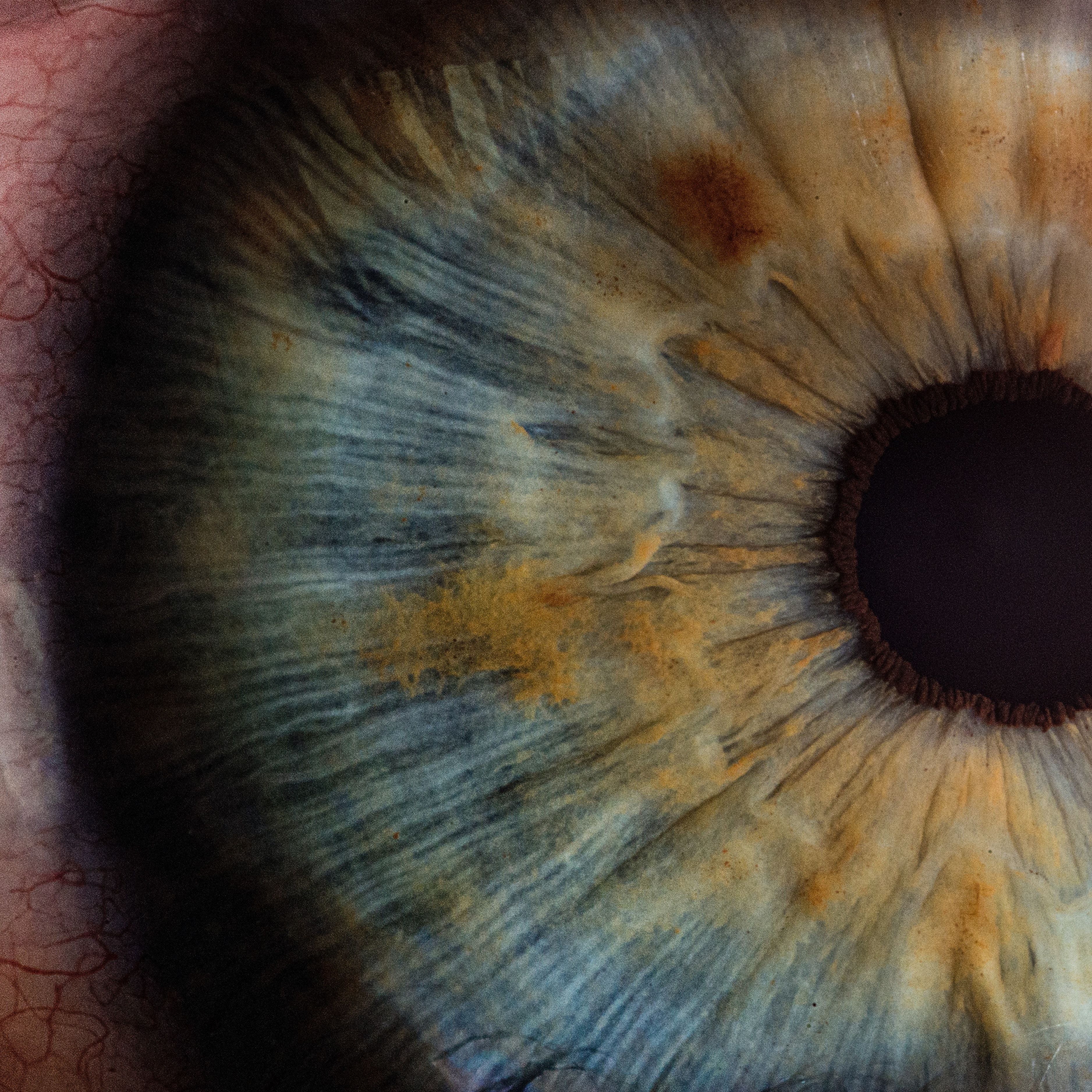
Dr. Brown discusses the most common conditions of patients treated for ocular trauma and highlights the range of performed procedures in the ED.
In an interview with HCPLive, Susannah Brown, MD, Assistant Professor of Ophthalmology, Medical University of South Carolina, discussed her specialty in ophthalmic plastic and reconstructive surgery and her role as the director of the Ocular Trauma service.
Her work involves both cosmetic and functional surgeries of the eyelid, eye socket, and tear drainage system. As the director, Brown sees all patients that come in through the emergency room and inpatients in the hospital with any and all eye complaints.
“I think there's a misconception that the majority of eye trauma occurs in high risk jobs, things like that, when the reality is that the majority of eye trauma that we see occurs during household activities,” Brown said.
Brown went on to describe other conditions that bring patients into her care, including strokes, glaucoma, or macular degeneration. Often, patients with these medical conditions may be asymptomatic and a sudden change in comfort of vision brings them to the emergency room.
Within the emergency room, Brown highlighted the broad range of procedures performed. This included fractures of the eye socket, often from motor vehicle accidents or assaults, or procedures of the eyelid, such as a canthotomy, cantholysis or eyelid lacerations.
She further highlighted perforating injuries to the eye, an open or ruptured globe injury, which can be devastating for patients and require immediate emergency surgery.
“Sometimes despite our best efforts, these patients don't have vision that we're able to restore, but, often they do,” Brown said. “Those are always really rewarding or exciting cases, when we're able to take the patients to the OR, restore their acute trauma, and usually over several weeks following that time, help them recover discomfort and also vision from the accident.”