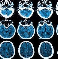
Those Deep Gray and White Matter Images Are Important in Multiple Sclerosis
MR-PET imaging can reveal inflammation and neurodegeneration in the brain, among other things, and a recent study showed that it can also help determine the progression of multiple sclerosis (MS).


MR-PET imaging can reveal inflammation and neurodegeneration in the brain, among other things, and a recent study showed that it can also help determine the progression of multiple sclerosis (MS).
Elena Herranz, PhD, from the Athinoula A. Martinos Center for Biomedical Imaging in Massachusetts, presented the findings at the 31st Congress of the European Committee for Treatment and Research in Multiple Sclerosis (ECTRIMS 2015) in Barcelona, Spain.
Previous research has identified that diffuse inflammation in the cortex, along with deep gray matter (DGM) and normal-appearing white matter (NAWM), may indicate MS progression. Herranz and her team aimed to explore the association more closely.
“Diffuse inflammation through activation of microglia and macrophages is thought to play a major role in determining diffuse demyelination and axonal damage in BM and NAWM,” Herranz explained during the presentation. “MRI can only indirectly detect inflammation in MS.”
The study compared the brain matter of 17 patients with MS (11 with secondary progressive multiple sclerosis, SPMS, and six with relapsing-remitting multiple sclerosis, RRMS) with healthy controls matched for age and sex. They used the 11C-PBR29 expression of the translocator protein (TSPO) to activate the microglia and macrophages. The participants underwent a 90-minute MR-PET scan that revealed substantial differences between the groups.
But is there a relationship between inflammation, neurodegeneration, Expanded Disability Status Scale (EDSS), and Symbol Digit Modalities Test (SDMT) scores? There indeed was an association between a higher EDSS score and greater standardized uptake value (SUV) in the thalamus, NAWM, and hippocampi. Only the patients with SPMS showed a relationship with lower SDMT scores and higher SUV scores in cortical and NAWM.
The 3T MR-PET data processing showed that compared to healthy controls, patients with MS had:
- 20% increase in white matter
- 10% increase in white matter lesions
- 50% increase in DGM in the thalamus
The amount of 11C-PBR28 in the whole cortex was 19% higher in those with MS. Further observation revealed that there was a significant decrease in cortical thickness in patients versus healthy controls. That cortical thinning correlated with the amount of inflammation. In addition, the measure of white matter in the thalamus was linked with the prominence of inflammation.
While only six of the patients with MS had cortical lesions, there were 51% observed than in healthy controls. Thalamic volume, however, did not appear to have any correlation with disease activity.
“Diffuse increase in PBR28 uptake was associated with neurodegeneration,” Herranz concluded. The team is currently working on finding the “correlation between inflammation and structural MRI data.”


