Unilateral Rash on the Back of a Mature Woman
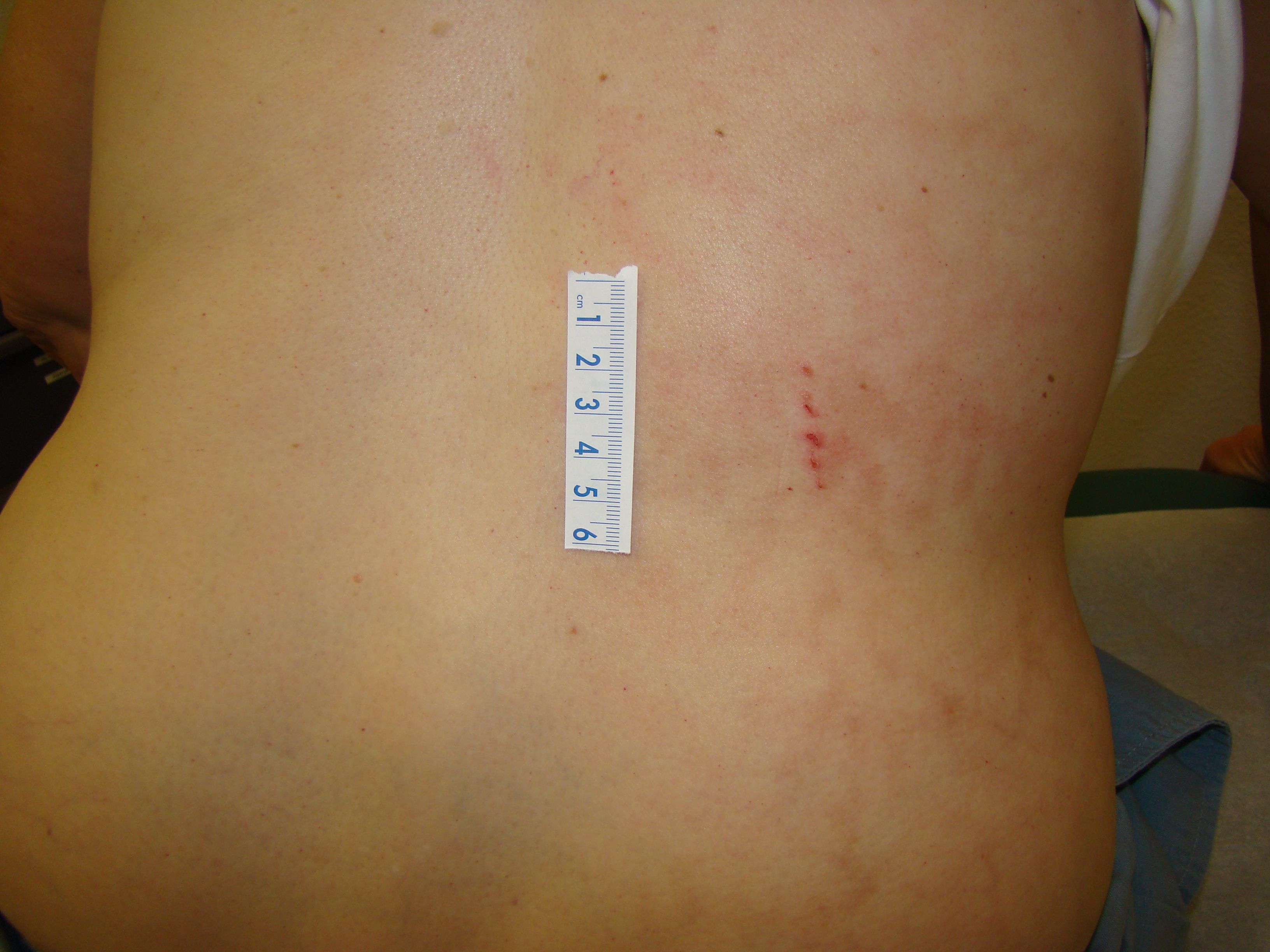
This 70-year-old woman noticed a rash one week ago in her right lumbar area with mild pain and change in skin color.
This 70-year-old woman noticed a rash one week ago in her right lumbar area with mild pain and change in skin color. The day prior to evaluation she was diagnosed with shingles and started on acyclovir. Her medical history is significant for shingles in the past and she was immunized after the event. She also has multiple sclerosis, bipolar disorder, spondylolisthesis and fibromyalgia with low-level positive ANA and p-ANCA. Her treatments have included gabapentin, high dose prednisone, physical therapy, topical OTC ointments and the use of a heating pad for her back pain.
What is your diagnosis?
- Herpes zoster
- Erythema ab igne
- Livedo reticularis
- Striae
- Vasculitis
Diagnosis

Erythema ab igne is the result of chronic and repeated exposure to a heat source. Clinical findings include a reticulated, erythematous or hyperpigmented dermatosis, also described as a “mesh-like or net-like” rash in the exposed area. Currently, common causes for this condition include heating pads, hot water bottles and more recently laptop computers. It is usually only a cosmetic concern, self limited and carries a good prognosis, however it could increase the risk of squamous cell or merkel-cell carcinomas. Treatment includes identifying and avoiding further exposure, topical retinoids, hydroquinone, or 5-fluorouracil can be used for the hyperpigmentation.
Herpes zoster, commonly called shingles, typically occurs in a dermatomal distribution with burning pain preceding the vesicular eruption. The vesicles evolve into pustular or hemorrhagic lesions that crust and resolve within 3 to 4 weeks. Prodromal malaise, headache and fever, are possible. This patient has a linear excoriation but no vesicles and hyperpigmentation would be a late feature of zoster.
Livedo Reticularis is characterized by a purplish mottled pattern of discoloration, with reticulated cyanotic areas surrounding paler central cores. Described as a “fishnet” pattern, it can be either idiopathic or a manifestation of a systemic disease. In idiopathic cases it is usually an asymptomatic cosmetic concern, as opposed to secondary cases in which it is usually related to rheumatologic diseases mainly antiphospholipid syndrome.2 In contrast, this patient's rash is localized to the area where she was using a heating pad for pain.
Striae are the consequence of normal skin stretching with subsequent rupture of elastic fibers in the reticular dermis. Initially the striae are erythematous and over time fade but are rarely hyperpigmented. These occur in regions of chronic tension such as the abdomen, areas of adipose tissue or edema, shoulders, thighs or breasts. Common clinical scenarios include abdominal distension from pregnancy, obesity or ascites. Cushing’s syndrome, with excess cortisol, weakens the dermis causing striae under normal tension.
Dermatologic manifestations of vasculitis have a broad presentation that must be correlated clinically along with laboratory findings. These include livedo reticularis, palpable purpura, nodules, ulcers and gangrene. Small-vessel vasculitis is often associated with palpable purpura of the lower extremities, while medium-vessel vasculitis causes nodules, ulcers and gangrene. This patient had rheumatologic markers that were positive, but only at low levels and had no correlation with the current physical exam.
References:
[1] Miller, Kristen; Hunt, Raegan; Chu, Julie; Meehan, Shane; & Stein, Jennifer. (2011). Erythema ab igne. Dermatology Online Journal, 17(10). Retrieved from: http://escholarship.org/uc/item/47z4v01z
[2] Usatine RP, Smith MA, Chumley HS, Mayeaux EJ, Jr... Chapter 200. Erythema Ab Igne. In: Usatine RP, Smith MA, Chumley HS, Mayeaux EJ, Jr... eds. The Color Atlas of Family Medicine, 2e. New York, NY: McGraw-Hill; 2013
[3] Usatine RP, Smith MA, Chumley HS, Mayeaux EJ, Jr... Chapter 124. Zoster. In: Usatine RP, Smith MA, Chumley HS, Mayeaux EJ, Jr... eds. The Color Atlas of Family Medicine, 2e. New York, NY: McGraw-Hill; 2013
[4] LeBlond RF, Brown DD, DeGowin RL. Chapter 6. The Skin and Nails. In: LeBlond RF, Brown DD, DeGowin RL. eds. DeGowin's Diagnostic Examination, 9e. New York, NY: McGraw-Hill; 2009
[5] Hellmann DB. Chapter 29. Introduction to Vasculitis: Classification & Clinical Clues. In: Imboden JB, Hellmann DB, Stone JH. eds. CURRENT Rheumatology Diagnosis & Treatment, 3e. New York, NY: McGraw-Hill; 2013.
About the Author

Daniel Stulberg, MD, is a Professor of Family and Community Medicine at the University of New Mexico. After completing his training at the University of Michigan, he worked in private practice in rural Arizona before moving into full-time teaching. Stulberg has published multiple articles and presented at many national conferences regarding skin care and treatment. He continues to practice the full spectrum of family medicine with an emphasis on dermatology and procedures.
Alberto Aguayo-Rico, MD, is a graduate of the Escuela de Medicina Ignacio A. Santos at the Tecnológico de Monterrey in Monterrey, Mexico and currently is a third year Internal Medicine Resident in the Primary Care Track at the University of New Mexico.
