Pulmonary Cavity, Lung and Skin Nodules, and Persistent Cough
Prepared by Charles L. Murphy, medical student, and Elena Kret-Sudjian, MD, Assistant Professor, Division of
Pulmonary and Critical Care Medicine, University of California Davis School of Medicine, Sacramento, Calif
Coccidioides immitis
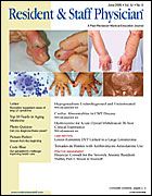
A 49-year-old, previously healthy woman from Bakersfield, California, presented with fever, chills, and a nonproductive cough. Three weeks earlier she was treated for community-acquired pneumonia, but her symptoms continued and worsened, prompting hospitalization. Physical examination revealed 2 small, tender nodules on one thigh. Chest x-ray showed a left-upperlobe infiltrate with possible cavitation (Figure 1). Chest computed tomography (CT) showed a left-upper-lobe cavity adjacent to the pleura, along with innumerable, bilateral, small lung nodules (Figure 2). Test results for sputum acid-fast bacillus and HIV infection were negative. A serum complement-fixation titer was also negative, but serum immunoglobulin M antibodies were positive. This serological finding along with positive culture from skin nodule biopsy established the diagnosis of coccidioidomycosis. Treatment with fluconazole resulted in resolution of her symptoms.
Points to Remember: Consider coccidioidomycosis in patients from endemic areas who have persistent respiratory symptoms. Endemic areas are the central valley of California, the desert southwest, and parts of Mexico and South America.
Diagnosis: Disseminated coccidioidomycosis.