Oral Spots with Anemia
Prepared by Grant E. Lattin, Jr, MD, Resident, William T. O
'Brien, Sr, DO, Resident, and Darick L. Jacobs, MD, Chief,
Interventional Radiology, Department of Radiology, David Grant US Air Force Medical Center, Travis Air Force Base, Calif
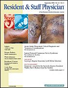
A69-year-old man underwent evaluation for anemia. Other than epistaxis requiring surgical clips, his medical history was unremarkable. Physical examination revealed telangiectases on his tongue and buccal mucosa (Figure 1), as well as on his palms and soles. The patient said that most of his family members had similar lesions. Colonoscopy identified a mass in the descending colon, from which pulsatile bleeding occurred when a biopsy was attempted. Angiography showed that the mass was a large, arteriovenous malformation (Figure 2); similar lesions were evident in the liver. These findings were diagnostic of hereditary hemorrhagic telangiectasia (HHT), also known as Osler-Weber-Rendu disease.
Points to Remember: HHT is an autosomal dominant
inherited disorder characterized by vascular lesions that variably appear in the nose, skin, brain, lung, or gastrointestinal tract. As seen in this patient, large arteriovenous malformations involving major organs may occur.
Diagnosis: Hereditary hemorrhagic telangiectasia.