An Irregular Visit to the Doctor
A 75-year-old woman with a history of coronary artery disease, hypertension and hyperlipidemia presents with shortness of breath with exertion.
A 75-year-old woman with a history of coronary artery disease, hypertension and hyperlipidemia presents with shortness of breath with exertion. She is normally very active.
When she is resting, she has no symptoms. But upon walking short distances she becomes easily winded. Nurse intake reveals a heart rate of 110 beats per minute and blood pressure of 110/65. She has a normal lung exam but her cardiac exam reveals a mildly irregular rhythm with a midsystolic murmur at the right upper sternal border. An EKG is performed.

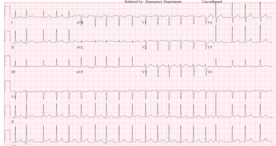

The EKG demonstrates a narrow complex rhythm with a relatively fast rate (approximately 120 bpm) and appears irregular. The irregularity is most evident at the left and right areas of the EKG. However, a quick check with the calipers will demonstrate that there is no consistent interval between QRS complexes throughout the EKG. Furthermore, there is no consistent P wave seen in the EKG. The patient above has atrial fibrillation with rapid ventricular rate with nonspecific ST and T wave abnormalities.
At the right side of the rhythm strip in V1, there appears to be flutter-like waves between beats 3rd to 4th from the right. Because V1 is closest to the atria, atrial activity can often be seen in this lead. The fibrillatory waves can be fine or coarse. Sometimes coarse fibrillatory waves can be mistaken for flutter waves, but the atrial activity associated with fibrillation will be inconsistent in morphology. Another rhythm in the differential would be multifocal atrial tachycardia. This would have 3 or more defined P wave morphologies and is often associated with hypoxia.
The ventricular response is typically irregular. But there are situations when it may be regular including in the setting of complete heart block from AV nodal disease or from digitalis toxicity. If the ventricular rate is less than 100 bpm, consider some underlying conduction disease before committing to a treatment regimen.