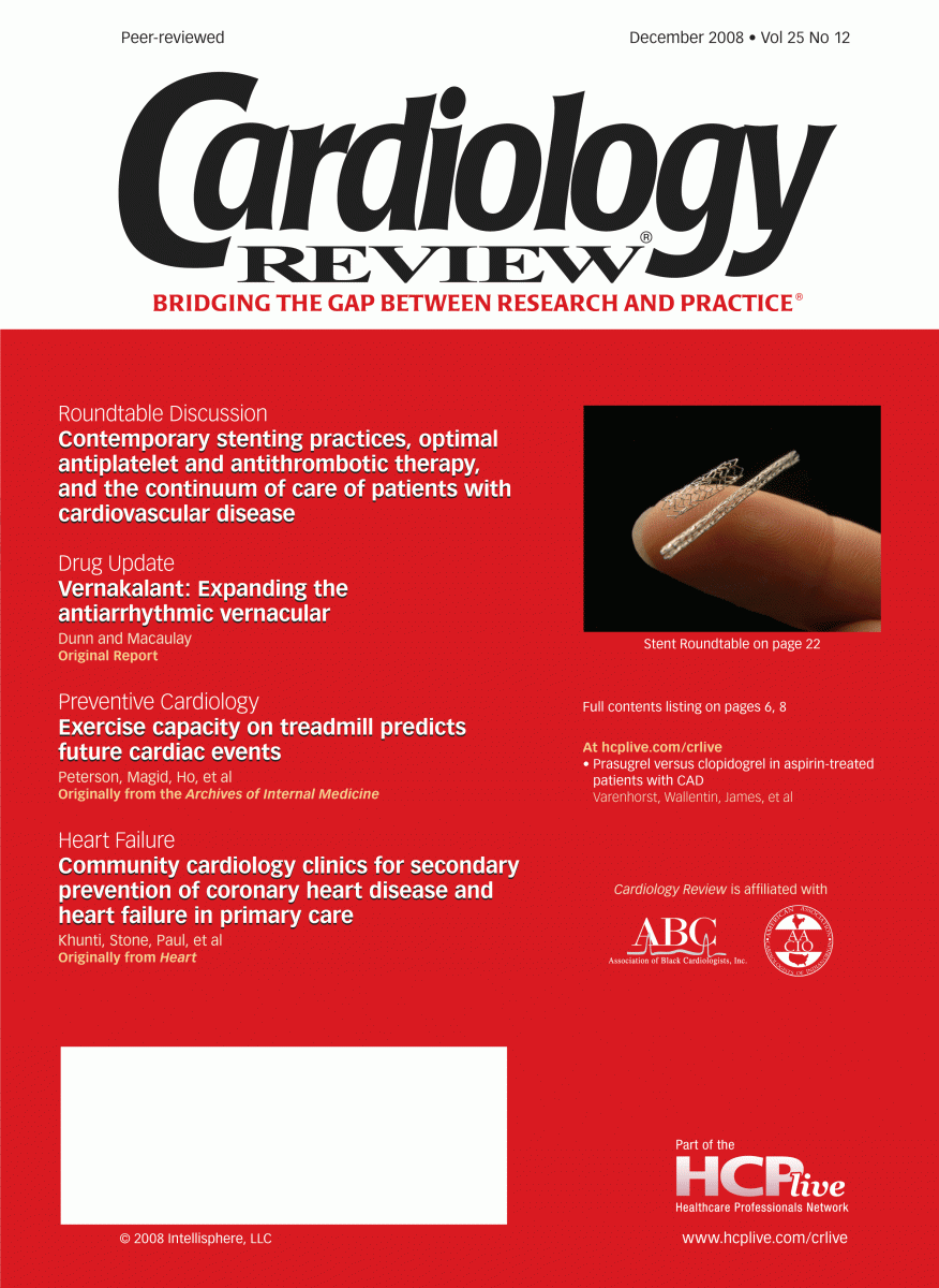
Contemporary stenting practices, optimal antiplatelet and antithrombotic therapy, and the continuum of care of patients with cardiovascular disease: A roundtable discussion
The first stent was invented in 1969 by Charles Theodore Dotter, experimenting on canine peripheral arteries.
The first stent was invented in 1969 by Charles Theodore Dotter, experimenting on canine peripheral arteries. Eight years later, the first coronary angioplasty was performed by Andreas Gruentzig in Switzerland, and in 1986, the first human coronary heart stent was implanted by Jacques Puel in France. Since then, there have been considerable technological advances in percutaneous coronary intervention (PCI), resulting in improved cardiovascular outcomes. One of the most significant developments to date is the advent of drug-eluting stents (DESs). This roundtable discussion will provide insights on currently available first- and second-generation DESs; examine advantages and disadvantages of DESs versus bare-metal stents (BMSs); review optimal antiplatelet and antithrombotic regimens during and after PCI; discuss imaging advances in detecting coronary artery disease (CAD); and look at strategies that might help to further improve the care of patients with cardiovascular disease (CVD). We have assembled an august faculty of cardiovascular experts to provide practical clinical insights into these issues. —
Debabrata Mukherjee, MD
DESs: Stent thrombosis, restenosis, and cardiovascular outcomes
D. Mukherjee: What mechanical strategies during PCI can be used to mitigate the risk of stent thrombosis in patients receiving DESs?
S. Sharma: DESs have mostly been compared with BMSs in the early time period following deployment (6 months to 1 year), and the rate of stent thrombosis seems to be similar during this time. It is only after 1 year when the issue of late stent thrombosis arises, which appears to be significantly higher with DESs. Early stent thrombosis may occur because of inadequate stent expansion and edge dissections. Therefore, optimization of stent deployment at the time of intervention will help mitigate the risk of early stent thrombosis.
How can we achieve this? One strategy, which has been widely used and reported in the literature, is intravascular ultrasonography (IVUS). This modality will help interventionalists select the right stent size, ensure full stent expansion and that the stent is touching the media, and resolve the issue of incomplete stent apposition, all of which could result in early stent thrombosis. If a thrombus is identified in the lesion, thrombectomy using an AngioJet® or another device should be undertaken. In cases of acute myocardial infarction (MI), removing the thrombus and placing a DES may translate into a lower stent thrombosis rate at follow-up. In cases of thrombus due to improper stent expansion or when there are calcified lesions that prevent the stents from fully expanding despite being deployed at a very high pressure, adjunct lesion modification using a cutting balloon or rotational atherectomy prior to stent deployment will help achieve good stent expansion.
This in turn will translate into getting a better lumen at the end of the procedure, and, subsequently, lower rates of early and late stent thrombosis. Another issue is incomplete stent apposition, where the stent does not come into contact with the media, resulting in a gap behind the stent struts. In these cases, once antiplatelet therapies are stopped, which traditionally has been after 1 year, a thrombus may develop behind the stent struts because there is no intimal hyperplasia in the DES, leading to late stent thrombosis. This scenario rarely happens with BMSs because there is intimal hyperplasia, which fills up the gap behind the stent struts.
Patients with acute coronary syndromes (ACSs) and MI will have higher late stent thrombosis rates because there is usually a thrombus behind the stent struts.
D. Mukherjee: Do you routinely use IVUS in your practice or are there angiographic characteristics that you use to determine if a patient needs IVUS? Also, do you routinely postdilate your DESs or do you deploy them at a high pressure? If you’re not postdilating them, at what pressure do you deploy them?
S. Sharma: Let’s talk about IVUS first. By 2004 and 2005, DESs were on the market with a 90% or more adoption rate, yet the use of IVUS during PCI varied from country to country. In Japan, for instance, over 80% of cases received IVUS, but in the United States that number was somewhere around 12% to 13%. When the issue of late stent thrombosis came to light in 2006, there was a big boost in IVUS use. Currently, we use IVUS in about 30% of stent deployments at our practice. IVUS is really helpful for stent optimization, but do you need to use it in every case? Probably not, but it may help. Much of the available X-ray equipment has a “stent boost” program that allows you to see the stent, whether it’s fully expanded or not. If you see incomplete expansion, you can go back and dilate with the balloon. At Mount Sinai, we probably use IVUS with a DES in about 25% to 30% of patients. IVUS doesn’t have to be used in everyone, but definitely in more complex cases, such as multiple stent placements, bifurcations, or stenting for left main CAD, which are high-risk groups for subsequent stent thrombosis. As for optimal dilatation pressure and postdilatation pressure, our practice has been a little different. Many people routinely postdilate all DESs. Until the availability of new-generation DESs—Medtronic’s Endeavor® and Abbott Vascular’s XIENCE™—when we used CYPHER® and TAXUS®, we used to just take their stent balloon to about 18 to 20 atm. In about 8% to 10% of cases, we still saw the stent incompletely expanded, or based on the IVUS findings, we went ahead with a high-pressure, noncompliant balloon dilatation of 1:1 vessel size and expanded the stent at 20 to 24 atm. This practice completely changed with the advent of the XIENCE and Promus™ stents, which have very thin struts and a very compliant balloon. What we learned when we started deploying these stents at 12 to 14 atm was that the stent really grows because of the compliance of the balloon. We’ve since changed our practice and now deploy the XIENCE between 8 and 12 atm, remove the stent balloon, and then use a high-pressure balloon that is shorter than the stent length to dilate the middle of the stent, or the area that has not been fully expanded, and take it to 20 to 22 atm.
R. Sharma: Are you going back with the strategy of routine high-pressure postdilatation with a noncompliant balloon?
S. Sharma: I would say in approximately 85% to 90% of cases. In a few cases, when we deployed a 2.5-mm XIENCE stent at 8 atm in a 2.25-mm vessel, our stent boost indicated that the stent was fully expanded and angiographically looked good. In those cases, I didn’t go back and dilate that stent, and we just left it. We actually did a few of these with IVUS, and the stent is nicely expanded at 8 atm in small noncalcified vessels.
R. Sharma: This is a very interesting observation. It will be interesting to see long-term results with this strategy using “stent boost.” We’re routinely deploying DESs at nominal pressures followed by high-pressure balloon inflations.
