
|Videos|January 29, 2021
Diagnosing and Assessing AD in Patients With Skin of Color
Author(s)HCP Live
Advertisement
Episodes in this series


Transcript:
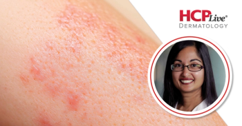
Andrew Alexis, MD, MPH, FAAD: Jamie, why don't you tell us about some of the challenges that a lot of health care providers might have in diagnosing atopic dermatitis in skin of color?
Jamie Weisman, MD: Candrice and Heather have done a great job telling us how hard it can be to recognize inflammation in skin of color and what can just look like color changes but is actually erythema. That is harder to appreciate. And this comes up in clinical trials too, where patients may have their disease underestimated, or resolution of the disease is in view because all you have is hyperpigmentation. The skin color hasn't normalized, but the inflammatory process has resolved. And those patients may be seen as treatment failures when they're not. They have a disease layered on top of their primary disease.
And ultimately, something we've been discussing earlier before we began this session was asking your patients. It's important to ask patients what their skin feels like. It may look fine to you; it may be deeply excoriated, and then you know it's itchy. But I find that if we don't dive in there, we don't have a real appreciation for the impact of the disease on the patient. What may not look like terribly inflamed skin, as Candrice alluded to, could be very itchy, could be uncomfortable for our patients. Unless we open the door for them to express that, we may never hear it.
Andrew Alexis, MD, MPH, FAAD: Well said. And I want to expand on one of the comments you made about the challenges, differentiating hyperpigmentation from erythema in the background of richly pigmented skin. As you pointed out, it's a double-edged sword. You can underestimate the severity of the disease if you aren't able to perceive the erythema accurately. And, on the other hand, you may underestimate the response to treatment or inaccurately consider someone a treatment failure if you can't differentiate the post-inflammatory hyperpigmentation from active lesions.
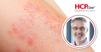
Do you agree with that, Jamie?
Jamie Weisman, MD: Yes, absolutely, that's a dilemma. And, again, I it's important to get your patient's perspective. If they have resolution of itch, there's a good chance they have resolution of the underlying inflammatory process. It's also very tactile. Candrice alluded to the idea that you may not even be able to appreciate some of these follicular-based lesions unless you actually touch the patient. And, again, you could really appreciate warmth and inflammation—that's the old rules of inflammation, tumor, rubor, dolor, calor—but you have to be willing to touch the patient. Talk to the patient, physically touch the patient. I think you're going to get a better idea of what's going on if you do those things.
Andrew Alexis, MD, MPH, FAAD: Exactly. Palpation, side lighting, history, all of that can help clinch the diagnosis and accurately assess the severity of atopic dermatitis. I would just add that when it comes to color, I like to teach my residents and other colleagues to sort of broaden the definition of erythema in a richly pigmented-skinned individual. Don't just look for red. There can be shades of red, brown, purple, gray. You sort of must broaden your palette if you will, when thinking about what redness looks like in the background of darkly pigmented skin.
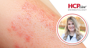
Heather, do you have any recommendations for accurately assessing atopic dermatitis in skin of color populations?
Heather Woolery-Lloyd, MD: When we do studies in clinical trials, they tell us typically to grade up one. If you would normally grade that level of erythema in a white patient as a 1, that same visual look, you should grade up because we do know that the skin color and pigmentation causes us to underestimate the degree of erythema. That's just a little clinical pearl that I got from clinical trials that visually what we see is actually 1 grade more severe when you're looking at it in a skin of a patient of color.
Andrew Alexis, MD, MPH, FAAD: Something I like to do is I like to calibrate. I like to teach that calibrating your eyes is a good approach, in that you first look at the patient's uninvolved nonlesional skin, and then you understand what their baseline is. And then you look at the lesion, and you sort of do a delta in your mind. And with that, then you can begin to appreciate the color change more accurately.
Let's talk about comorbid conditions. We all know that atopic dermatitis can be associated with other disorders that are Th2 driven. Can you speak to any issues that are relevant to skin of color populations in comorbid conditions? Heather, why don't you take that?
Heather Woolery-Lloyd, MD: As I mentioned earlier, atopic dermatitis is more common and more severe in Black children. And, interestingly, the comorbid conditions that we typically see with atopic dermatitis are asthma and allergic rhinitis. And, not surprisingly, Black children also have higher rates of asthma, and sadly, they're more likely to be hospitalized for their asthma and actually 10 times more likely to die from complications of asthma.
As we see in atopic dermatitis, it's more common and more severe in Black children and we see that same comorbidity. Black children with atopic dermatitis are more likely to have asthma. And Black children in general who have asthma are more likely to be hospitalized and more likely to die from complications of asthma.
Andrew Alexis, MD, MPH, FAAD: Really staggering statistics then. Thank you for making that point.
Transcript Edited for Clarity
Advertisement
Related Content
Advertisement


Latest CME


Advertisement
Advertisement
Trending on HCPLive
1
Risankizumab Versus Icotrokinra: New Data on Adults with Moderate-to-Severe Psoriasis
2
How Well-Being in Psoriasis is Being Assessed Via WHO-5, With Ulrich Mrowietz, MD
3
FDA Approves Atacicept (Trutakna) for IgA Nephropathy
4
Q2 2026 Recap: Gastroenterology News & Updates
5