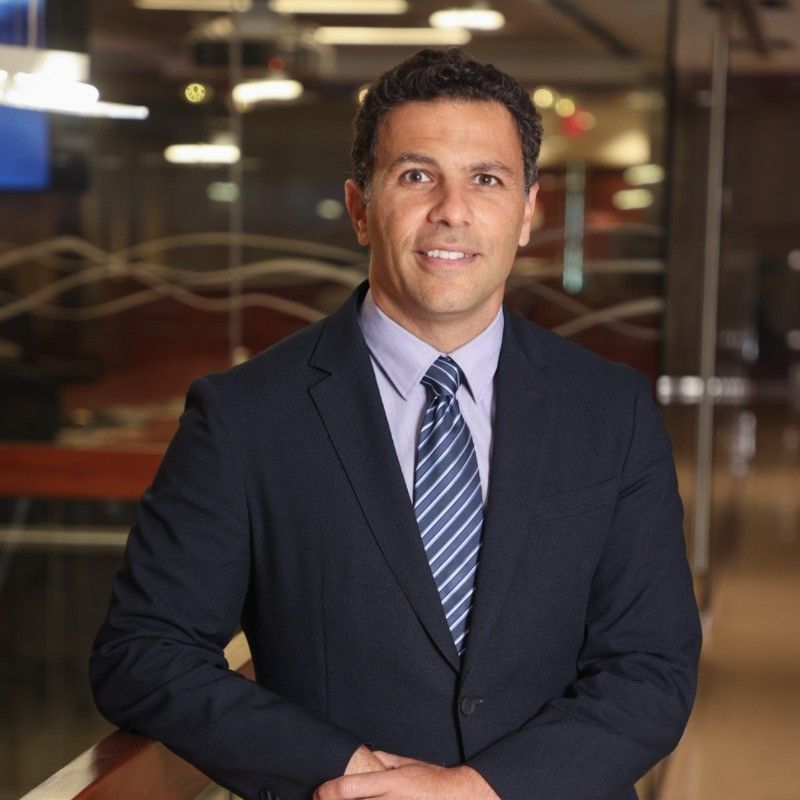
Discussing Advances in Structural Heart Disease, with Elliot Elias, MD, MPH
Elliot Elias, MD, MPH, provides perspective on recent advances in structural heart disease as well as highlighting the upcoming 40th annual Echocardiography and Structural Heart Symposium at Baptist Health.
Elliot Elias, MD, MPH
Credit: LinkedIn

The management of structural heart disease has undergone significant changes in the last decade. With the growing popularity and indications of transcatheter aortic valve replacement (TAVR) as well as imaging and other treatment modalities, education and dissemination of best practices for the management of structural heart disease have become a focus.
These topics will be the focal point of the 40th Annual Echocardiography and Structural Heart Symposium. A 2-day event hosted by Baptist Health Miami Cardiac and Vascular Institute, the event boasts more than 3 dozen sessions featuring expert presenters discussing topics including specific disease pathologies, the optimal role of imaging specialists on the cardiovascular care team, and pitfalls in contemporary management strategies.
With an interest in learning more about the event and its agenda, the editorial team of HCPLive Cardiology sat down with Elliot Elias, MD, MPH, moderator for the symposium and medical director of Cardiac and Structural Imaging at the Baptist Health Miami Cardiac & Vascular Institute. As part of the meeting, Elias will moderate or present on topics related to aortic stenosis, evaluation of prosthetic heart valves, evaluation of imaging in valvular heart disease, and management of mitral valvular disease regurgitation.
In the following Q&A, conversation, Elias provides an overview of several sessions included in the symposium, offers additional insight into contemporary trends in the management of structural heart disease, and discusses the need for symposiums such as those one for a field with the rapid. Level of advancement seen within structural heart disease.
Elliot Elias, MD, MPH, on Advances in Structural Heart Disease
HCPLive Cardiology: Why are symposiums such as this one necessary for a rapidly evolving space like structural heart disease?
Elias: I think it gives us a chance to really focus on the expertise we have at Baptist Health. We're at a tertiary center, almost a quaternary center, where we've recruited surgeons and cardiologists from all over the country. We've assembled a center where surgeons, cardiologists, radiologists, and anesthesiologists collaborate to perform complex cases for patients with complex diseases. The symposium provides us with an opportunity to spotlight the individuals who perform these procedures. About 80% of the conference features actual cases from Baptist Hospital, allowing us to integrate imaging complexities into patient care.
Another great aspect of this symposium is our ability to give back to the community from both a cardiology and surgery perspective, focusing on the conference attendees, primarily general cardiologists and stenographers. This symposium stands out because most others concentrate on one or the other, catering to either personal stenographers or cardiologists. However, this forum blends both, creating a unique environment where attendees can explore imaging comprehensively.
From my perspective, stenographers are the first point of contact with the patient. They guide patients from the waiting room to the examination bed, and then utilize echocardiograms to diagnose diseases. They form the backbone of cardiology and structural imaging, and without their expertise, we wouldn't be where we are today. Our approach involves examining everything through their eyes, appreciating the patient's journey from the waiting room, and valuing their expertise. We also delve into how doctors utilize this imaging, interpret it, request additional imaging, and communicate with the intervention team, including structuralists and surgeons. This collaborative effort allows patients to receive treatments that were not available just 15 years ago.
HCPLive Cardiology: Could you dive into how the role of stenographer has evolved in recent years?
Elias: So, I think that the Baptist healthcare system and amongst all the other leading imaging centers around the country, the stenographer really is at the base of this, and we have constant interaction with them. They need to have constant interaction with us to really feel like they're part of making that diagnosis. We really tried to stay away and change the idea that it's just going to be someone that's in a room, taking pictures, and, then, sending those pictures to a screen where the doctors in another room are reading the pictures and then making the diagnosis, and to push more for an interactive approach where they are asking questions and we are asking them to actually do more, and almost being more of a diagnostician rather than just the person that is getting images.
HCPLive Cardiology: What type of role could artificial intelligence (AI) serve in management of structural heart disease?
Elias: Artificial intelligence is the future, how we integrate that in our practice is important. We have regular meetings looking for the best ways to integrate and looking for the best software as well as the best imaging solutions for our practice at Baptist Health and for our group as a whole. When we start looking at the specifics around AI, such as what does AI look like for imaging, the tools that are out there are pretty impressive. They're not ready at this point in time, but some of the models are really.
When you start looking at it from the stenographer’s standpoint, telling them how to angle it ever so slightly, so they can get optimal imaging is pretty impressive. The background to that is when a stenographer scans a patient, obviously there's no markers on the patient, as you know, we would put our ultrasound probe here, or we would angle it, you know, just a slight 10 degrees more that way. But artificial intelligence is actually going to look at the images that are being produced and tell the stenographer how to optimize the images by either rotating, angling, or positioning that probe in a different way so they can optimally obtain those images.
That's something that I think is going to be really game-changing for them because we're going to be able to image our patients efficiently and optimally. From the cardiologist’s perspective, we're going to have reports that are in a sense already going to say, or help us say, "Okay, we're going to have an alert this patient may need treatment sooner than later, this patient needs to be treated, or followed up with another imaging study within three months instead of six months. This patient should be seen in clinic at this time", and it really is going to integrate us all as a team from the stenographer standpoint, from a patient standpoint, and from the cardiologist standpoint in a way that is just simply not possible without artificial intelligence.