Hyperpigmented Back Lesion in a Young Woman
This 28-year-old woman presented with a large hyperpigmented patch on the right side of her upper back. Although the lesion developed over the past 2 years, it has been "sinking in" over the past 2 weeks. The patient denies any trauma, pain, discomfort, or pruritis, as well as any family history of cancers, lymphoma, or autoimmune disease. However, she has a history of gastroesophageal reflux disease and onychomycosis, and her grandfather has a similar lesion.
a) Becker’s nevus
b) Morphea, or localized scleroderma
c) Panniculitis
d) Extragenital lichen sclerosus
e) Mycosis fungoides, or cutaneous T-cell lymphoma
Diagnosis


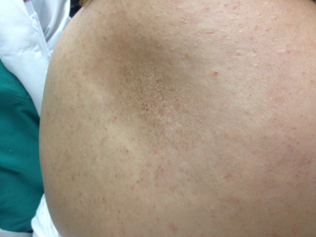
Morphea is a localized form of scleroderma that presents as a sharply demarcated, indurated plaque, which may be flat, slightly raised, or somewhat depressed. Usually, the lesion first appears with red or purple discoloration that eventually matures to brown or yellow-brown patches. It most often involves the trunk with no systemic inflammatory findings.1
In this case, the patient’s diagnosis was confirmed by a punch biopsy that revealed a sclerotic dermis accompanied by a sparse lymphoplasmacytic infiltrate consistent with morphea.
Morphea is most commonly treated with topical, intralesional, or systemic steroids, if needed. Calcipotriene, imiquimod, phototherapy, and methotrexate are additional options for the condition’s active inflammatory phase.2
Becker’s nevus also presents as an irregular hyperpigmented patch on the torso or upper arm in young adults. However, it predominantly affects males, often has hypertrichosis, and is sometimes thickened, but only rarely depressed.3
Panniculitis is an inflammation of the subcutaneous fat that usually causes erythematous tender nodules. The most common form of the disease is erythema nodosum, which predominantly affects women’s legs in contrast to this patient’s truncal hyperpigmentation and lack of tenderness.4
Lichen sclerosus presents as white, thickened plaques or atrophic patches typically in the genital and perianal areas. However, in the 15-20% of cases that involve the thighs, buttocks, breasts, shoulders, or neck, the condition is known as extragenital lichen sclerosis.5
Mycosis fungoides, the most common type of cutaneous T-cell lymphoma, can present in various forms, though it is usually eczematous or scaly. When the condition fails to respond to topical steroids and emollients, a biopsy is used to make the diagnosis.6
References
1. Fett NM. Morphea (localized scleroderma). JAMA Dermatol. 2013 Sep;149(9):1124. http://archderm.jamanetwork.com/article.aspx?articleid=1740691.
2. Firat Y, Sireci S, Yakinci C, Akarçay M, KarakaÅŸ HM, Firat AK, Kizilay A, SelimoÄŸlu E. Isolated preauricular pits and tags: is it necessary to investigate renal abnormalities and hearing impairment? Eur Arch Otorhinolaryngol. 2008 Sep;265(9):1057-60. http://www.ncbi.nlm.nih.gov/pubmed/18253743.
3. Danarti R, König A, Salhi A, Bittar M, Happle R. Becker's nevus syndrome revisited. J Am Acad Dermatol. 2004 Dec;51(6):965-9. http://www.ncbi.nlm.nih.gov/pubmed/15583590.
4. Chopra R, Chhabra S, Thami GP, Punia RP. Panniculitis: clinical overlap and the significance of biopsy findings. J Cutan Pathol. 2010 Jan;37(1):49-58. http://www.ncbi.nlm.nih.gov/pubmed/19708879.
5. Colbert RL, Chiang MP, Carlin CS, Fleming M. Progressive extragenital lichen sclerosus successfully treated with narrowband UV-B phototherapy. Arch Dermatol. 2007 Jan;143(1):19-20. http://archderm.jamanetwork.com/article.aspx?articleid=410360.
6. Nashan D, Faulhaber D, Ständer S, Luger TA, Stadler R. Mycosis fungoides: a dermatological masquerader. Br J Dermatol. 2007 Jan;156(1):1-10. http://www.ncbi.nlm.nih.gov/pubmed/17199560.
About the Authors


Daniel Stulberg, MD, is a Professor of Family and Community Medicine at the University of New Mexico. After completing his training at the University of Michigan, he worked in private practice in rural Arizona before moving into full-time teaching. Stulberg has published multiple articles and presented at many national conferences regarding skin care and treatment. He continues to practice the full spectrum of family medicine with an emphasis on dermatology and procedures.
Ahmed A. EL-Emawy, MD, is a graduate of the University of New Mexico Medical School and currently a Family Medicine Resident at the University of New Mexico.


 May Offer Over PPIs, with Adelina Hung, MD")


