Article
Virtual Breast Models Allow Clinicians to Hone Diagnostic Skills
Author(s):
Researchers created a virtual breast that can be used to practice elastography ultrasound skills and interpret the results.


A virtual breast model has been created and can allow clinicians to improve their cancer diagnostic skills, according to a poster presented at the IEEE Ultrasonics Symposium, held in Chicago from September 3-6.
Researchers from Michigan Technological University in Houghton, Michigan created a virtual breast model test to identify potential tumors throughout the body and breast. This virtual breast testing was created in a phantom lab in the public domain used for rigorous testing.
The test uses imaging to identify the stiffness of breast tissue, and can identify possible cancer areas because cancer tissues are very stiff. One analogy presented is that normal breast tissue is like a yolk in a fried egg, but not all of the readings are that clear.
“Depending on who does the reading, the accuracy can vary from 95 percent to 40 percent,” Jingfeng Jiang, PhD, biomedical engineer at the University, explained in a press release. “Forty percent is very bad—you get 50 percent when you toss a coin. In part, the problem is that ultrasound elastography is a new modality, and people don’t know much about it.”
The idea behind the virtual breast model was so clinicians could improve their accuracy in reading suspicious mammograms, as surgeons practice on simulators. Jiang was on the University of Wisconsin-Madison team which developed ultrasound elastography, and hopes that by applying virtual ultrasound elastography to virtual breast models and evaluating the results, clinicians will advance their skills in the field.
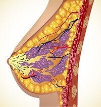
The model was generated from female cadaver, and included a variety of tissue types and anatomical structures like ligaments and milk ducts. Data from the Visible Human Project, which incorporates thousands of cross-sectional photos, was collected.
“Based on our preliminary study, the proposed virtual breast phantom can be used to perform sophisticated acoustic simulations and can also be used in quasi-static elastography applications,” the authors concluded.
In the future, the investigators hope to incorporate more anatomical structures to improve the realism of the virtual breast model and to make the phantom lab widely available for greater use for anyone that requires the training.