Publication
Article
Family Practice Recertification
White and Dark Lesions in a Newborn Infant
Author(s):
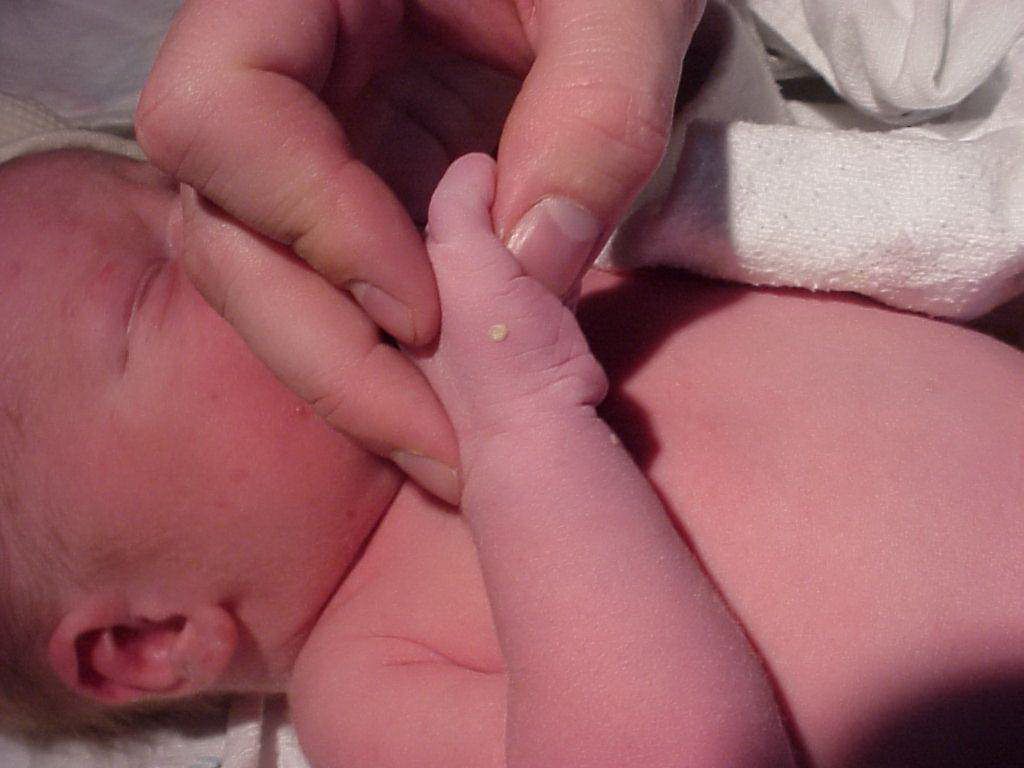
This newborn was found to have these fragile white lesions on the scrotum and extremities along with dark macules on the face. The child was otherwise asymptomatic and the pregnancy was uncomplicated, with no significant medication exposures and no family history of similar lesions.
This newborn was found to have these fragile white lesions on the scrotum and extremities along with dark macules on the face. The child was otherwise asymptomatic and the pregnancy was uncomplicated, with no significant medication exposures and no family history of similar lesions.
What is your diagnosis?
- Milia
- Miliaria
- Erythema toxicum neonatorum
- Herpes simplex virus
- Transient neonatal pustular melanosis
Diagnosis
Transient neonatal pustular melanosis is a benign condition affecting 5% of African-American infants and 1% of Caucasian infants. The name is very useful in remembering the nature of the disorder. The lesions start as short-lived pustular or vesicular lesions that leave behind hyperpigmented macules upon rupturing. The hyperpigmentation resolves spontaneously in several weeks and is completely asymptomatic. If the pustular process occurs in utero, the only signs of the disease are the hyperpigmented macules as seen on this infant’s face in the background. Pathology or a smear of the contents of the pustules reveals neutrophils without bacteria.

Milia present as white spots in 50% of newborns. They most commonly occur on the face due to small cysts of retained keratin. They resolve spontaneously with exfoliation of the upper layers skin. These lesions are not flaccid or easily ruptured like transient neonatal pustular melanosis and resolve without hyperpigmentation.1
Miliaria is also a disease of the newborn presenting in the crystallina form as small superficial clear vesicles with minimal surrounding erythema. It is due to obstruction of the sweat glands and when the obstruction is lower leads to miliaria rubra also called heat rash. In this second form as the name indicates there is erythema associated with the vesicular lesions. 1
In contrast to its alarming name, erythema toxicum neonatorum is a benign process seen in half of all newborns, most commonly on the trunk. The pustular appearing lesions are due to collections of eosinophils in contrast to the neutrophils of transient neonatal pustular melanosis and also in contrast, the lesions are surrounded by erythema.
Herpes simplex virus can cause life-threatening complications when contracted in utero or as a neonate. In utero infection is associated with preterm delivery, microcephaly, hydrocephalus and vesicular skin lesions. If it is due to vertical transmission at delivery, symptoms usually develop at one week with general signs of infection including lethargy, poor feeding, and may have cutaneous vesicles. In contrast this term infant was asymptomatic and appeared normal other than the skin lesions.
1.O'Connor NR, McLaughlin MR, Ham P. Newborn skin: Part I. Common rashes. Am Fam Physician. 2008 Jan 1;77(1):47-52.
3. Monteagudo B, Labandeira J, Cabanillas M, Acevedo A, Toribio J. Prospective study of erythema toxicum neonatorum: epidemiology and predisposing factors. Pediatr Dermatol. 2012 Mar-Apr;29(2):166-8
4. Rudnick CM, Hoekzema GS. Neonatal herpes simplex virus infections. Am Fam Physician. 2002 Mar 15;65(6):1138-42.
References:
About the Author

Daniel Stulberg, MD, is a Professor of Family and Community Medicine at the University of New Mexico. After completing his training at the University of Michigan, he worked in private practice in rural Arizona before moving into full-time teaching. Stulberg has published multiple articles and presented at many national conferences regarding skin care and treatment. He continues to practice the full spectrum of family medicine with an emphasis on dermatology and procedures.


