Article
Young People with Diabetes Show Significant Visual Improvement After PDR Surgery
Author(s):
However, the analysis suggests young adults with T2D who underwent vitrectomy for PDR had worse final visual acuity and more complications compared to those with T1D.


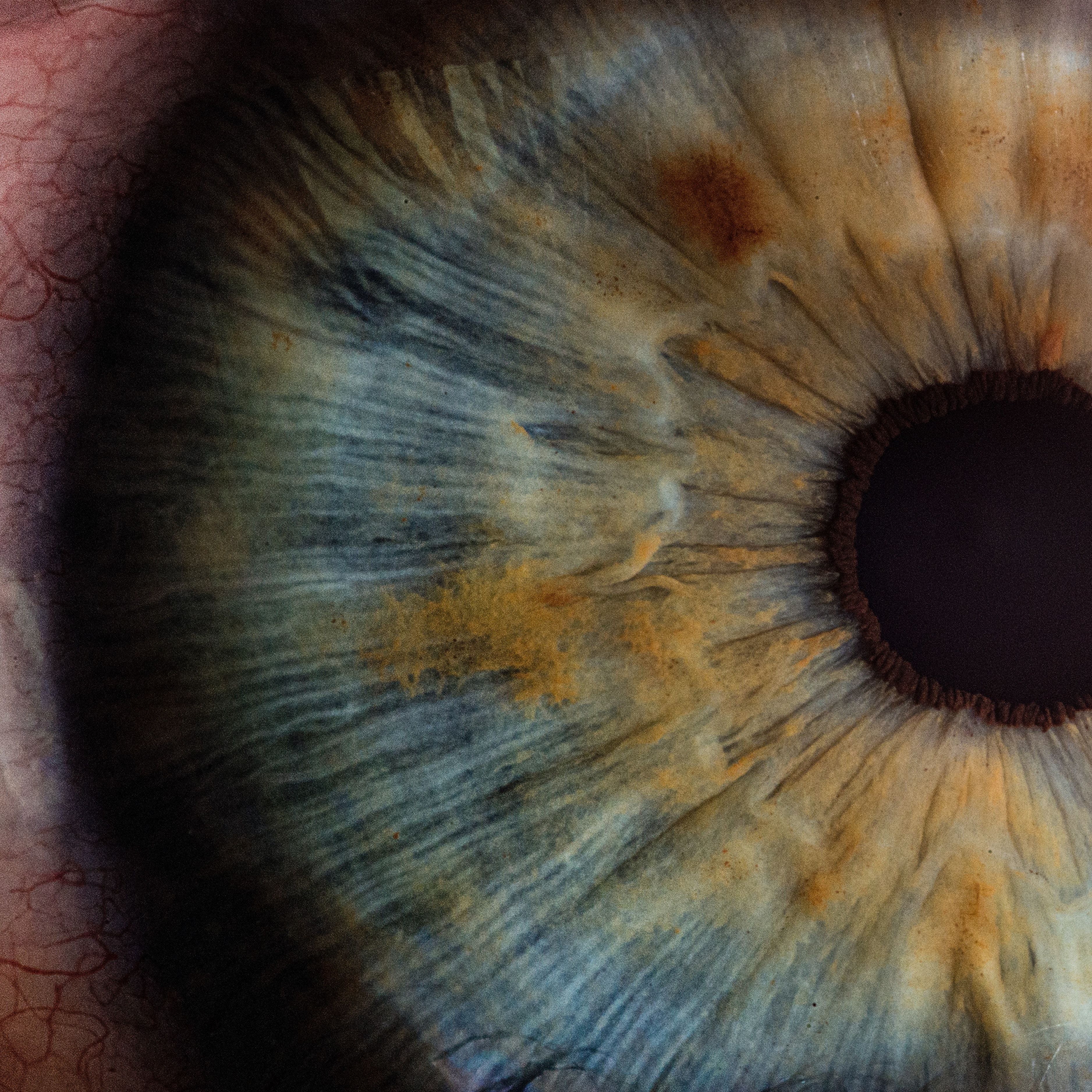
Credit: V2osk/Unsplash
Young adults with type 1 diabetes (T1D) or type 2 diabetes (T2D) with severe proliferative diabetic retinopathy (PDR) benefited from pars plana vitrectomy (PPV), experiencing a significant improvement in visual acuity.1
The retrospective results from China indicate, however, the T1D group had a greater percentage of patients in whom the postoperative visual acuity improved or remained stable, compared with those in the T2D group.
“These results suggest that PPV is effective in patients with severe PDR, with either T1D and T2D, and it can achieve significant visual improvement,” wrote the investigative team, led by Xin Huang, from the department of ophthalmology at Eye & ENT Hospital at Fudan University. “However, patients with T1D may have a better chance of visual improvement after PPV than patients with T2D.”
Prior literature has suggested young adults with T2D have double the risk of developing retinopathy and PDR sooner than young adults with T1D.2 Both the clinical manifestations and outcomes of vitrectomy for severe PDR in older have been reported, but there are little data on severe PDR in young adults, contrary to its growing prevalence in recent years. Huang and colleagues conducted a retrospective study to better understand the clinical characteristics and surgical outcomes of severe PDR in young adults with T1D or T2D.
The team reviewed the medical records of patients aged <45 years with T1D or T2D who underwent surgical treatment for severe PDR between January 2018 - July 2022. Indications for PPV included prolonged vitreous hemorrhage without absorption, tractional retinal detachment involving the macula, concurrent tractional rhegmatogenous retinal detachment (RRD), and severe active fibrovascular proliferation (FVP). Investigators retrieved detailed preoperative, intraoperative, and postoperative data from the medical records of this patient population.
Overall, the analysis enrolled 99 patients, comprising 140 eyes with severe PDR. A total of 24 eyes (18 patients) were included in the T1D group and 116 eyes (81 patients) were included in the T2D group. A much greater proportion of males was observed in both the T1D (77.8%) and T2D (65.4%) groups than females.
Those in the T1D group showed a longer duration of diabetes (P = .008), younger age at primary vitrectomy (P = .049), and a lower body mass index (P <.001), compared with the T2D group. Surgical records indicate the main indication of PPV in the T1D group was tractional RRD (11 eyes, 45.8%), while vitreous hemorrhage without other indications was more prevalent in the T2D group (31 eyes, 26.7%). The T1D group showed a greater proportion of eyes with RRD (P = .040), but a smaller proportion with TRD (P = .027), compared with the T2D group.
Analyses showed patients with T1D generally had worse preoperative BCVA, but better postoperative BCVA than patients with T2D. At the end of follow-up, investigators found BCVA had improved significantly compared with the preoperative BCVA in both groups (all P <.001). Data showed the percentage of eyes in which the postoperative visual acuity improved or remained stable was greater in the T1D group than those in the T2D group (P = .045).
Investigators noted the final BCVA was improved or stable in 100% and 85.3% of eyes in the T1D and T2D groups, respectively, and decreased in 0% and 14.7% of eyes, respectively. Meanwhile, the overall incidence of postoperative complications was significantly greater in the T2D group compared with the T1D group (P = .045).
In an assessment of risk factors associated with the final visual acuity, investigators found preoperative BCVA was associated with worse visual acuity in both groups. For the T1D group, the duration of diabetes (P = .031) and preoperative FVP (P = .004) were significantly associated with worse visual acuity. In the T2D group, preoperative RRD (P <.001) and postoperative NVG (P <.001) were significantly associated with worsening.
Huang and colleagues underscored the significance of these findings, particularly regarding early detection and intervention in managing PDR in young adults. Results suggest, according to the team, that individuals with T2D need more vigilant postoperative monitoring in order to effectively manage complications.
“In future research, we plan to continue following up with both groups of patients and supplementing our sample size in order to conduct more robust and long-term research on this topic,” investigators wrote.
References
- Zhang M, Zhang J, Xu G, Ruan L, Huang X. Comparison of Clinical Profiles, Demographics, and Surgical Outcomes of 25-Gauge Vitrectomy for Proliferative Diabetic Retinopathy in Young Adults with Type 1 or Type 2 Diabetes. Diabetes Metab Syndr Obes. 2023;16:1967-1975 https://doi.org/10.2147/DMSO.S412157
- Patricia B, Andrew JB, David OH, Brian GM. Ocular sequelae in a population-based cohort of youth diagnosed with diabetes during a 50-year period. JAMA Ophthalmol. 2022;140(1):51–57. doi:10.1001/jamaophthalmol.2021.5052