Publication
Article
How Should I Manage This Man's Hyperthyroidism?
Author(s):
Though laypeople are sensitized to associating "radioactive" with "cancer," this is an unwarranted fear when considering radioactive iodine ablation for hyperthyroidism.

A 42-year-old African-American male presents with a 3-month history of nervousness, palpitations, and weight loss. Although he reports that he has watched his diet during that time, he expresses surprise that he has lost 15 pounds. Over the past 2 months, he has become increasingly anxious and aware of a more rapid heartbeat. His wife complains that he has insisted on keeping the windows open in the bedroom, despite her requiring an electric blanket because it has been so cold. His past medical history is notable only for hypertension controlled with hydrochlorothiazide. You suspect a diagnosis of hyperthyroidism.
What findings on physical examination would support this diagnosis?
Viewed in isolation, this man’s symptoms are not specific. However, the constellation of nervousness, palpitations, rapid heart rate, possibly unexplained weight loss, and heat intolerance strongly suggest thyrotoxicosis. Other symptoms that can accompany thyrotoxicosis include fatigue, diarrhea, and deceased libido. In premenopausal women, oligomenorrhea is a common symptom, as well.
Physical findings that would support a thyrotoxicosis diagnosis include tachycardia, fine tremor of extended fingers, brisk and exaggerated deep tendon reflexes, an enlarged thyroid gland with or without nodules, proximal muscle weakness, difficulty maintaining attention, and warm, moist skin. It is sometimes possible to observe lid lag, where the sclera is visible above the iris when the eyes quickly move from superior gaze to inferior gaze.
The man’s physical examination is notable for a blood pressure of 130/54 and a pulse of 110 bpm. His extraocular movements are full with mild lid lag. There is no evidence of exophthalmos, though a mild tremor with outstretched hands is noted. The patient’s thyroid exam reveals a mildly enlarged, nontender gland without bruit. What laboratory tests would you order?
Initial testing should assess thyroid-stimulating hormone (TSH) and unbound thyroxine (T4), as well as free T4 index (FTI), which assesses bioavailable T4. Most circulating T4 is firmly bound to protein, and the hypothalamic-pituitary-thyroid (HPT) axis tightly regulates secretion of T4, which serves to complete the feedback loop to the pituitary. Thus, in clinical thyroid disease, the relationship between circulating TSH and circulating thyroxine is reciprocal.
Laboratory testing reveals TSH of <0.02 mcIU/mL (n 0.3-4.7). His free T4 index is 24 pmol/L (n 4.5-10.5). What is the differential diagnosis?
The differential diagnosis in this patient includes toxic multinodular goiter, toxic adenoma, functioning thyroid metastases, and Graves’ disease, which is hyperthyroidism due to autoimmune thyroiditis. Factitious hyperthyroidism is a rare event that is a medical correlate of Münchausen’s syndrome. If the patient was a woman, then I would also consider struma ovarii, which is a thyroid hormone-secreting ovarian tumor.
There is a close link between Graves’ and Hashimoto’s disease, and autoimmune thyroid injury is common to both conditions. Some experts believe the initial phase of Hashimoto’s may be subclinical, so its presentation may be delayed until clinical hypothyroidism develops. On the other hand, some Graves’ cases will gradually appear to remit for months until permanent hypothyroidism develops. Therefore, functionally autoimmune thyroid disease behaves as a continuum from Graves’ to Hashimoto’s.
What diagnostic tests would you order next?
In this particular case, it would be reasonable to begin treatment without further testing. Most physicians would also obtain a complete blood count, liver profile, and electrolytes, including calcium with blood urea nitrogen and creatinine. While those tests do not contribute to diagnostic accuracy, they do screen for complications that might affect a treatment choice.
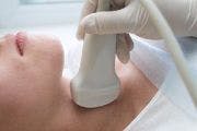
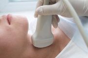
If the examiner is not completely content with the physical examination findings, then imaging the gland is necessary to distinguish among Graves’ and nodular disorders. In the setting of euthyroidism and a palpable nodule, ultrasound imaging is the appropriate test. However, radionuclide imaging is more appropriate in the case of hyperthyroidism.
Diagnostic options include sodium iodide 123 (I-123), technecium-99m (Tc-99m) sodium pertechnetate, or sodium iodide 131 (I-131). When it is easily available, orally administered I-123 is the preferred agent. Though intravenous Tc-99m sodium pertechnetate offers the lowest radiation exposure, it has the drawback of requiring an additional low-dose iodide scan to assess iodide uptake.
The Practice Guideline for the Performance of Thyroid Scintigraphy and Uptake Measurements collaboratively revised by the American College of Radiology (ACR), the Society for Pediatric Radiology (SPR), and the Society of Nuclear Medicine (SNM) strongly discourages iodide-131 for diagnostic use, since the dose of radiation is much higher.
The man’s thyroid scan reveals diffusely increased homogeneous tracer uptake with no identified nodules. Both lateral lobes of the thyroid appear enlarged. Uptake at 4.5 hours was 54.2% and increased to 75.5% at 24 hours. You decide this confirms the diagnosis of Graves’ disease. What are the treatment options?
Initial treatment should aim to control thyrotoxic manifestations with a beta-blocker (BB). Propranolol in 3-4 divided doses has been used in early studies, but once-daily 25-100 mg atenolol is more convenient.
There are 3 acceptable methods for the definitive treatment of hyperthyroidism: antithyroid medication, radioactive iodine ablation, and surgery.
#1. Antithyroid Medication
Propylthiouracil (PTU) and methimazole both inhibit thyroid peroxidase, so they decrease the oxidation and organification of iodine. PTU inhibits the peripheral conversion of T4 to triiodothyronine (T3), although the effect is only significant in thyroid storm. Methimazole is the preferred drug in most circumstances because it requires twice-daily doses, while PTU initially requires dosing every 6-8 hours.
Testing FTI every 3-4 weeks enables the recognition of stable dosing. It is important to understand that resetting the HPT axis takes months, so rely on FTI to assess short-term changes, rather than TSH. Adjusting the dose over time maintains a stable euthyroid state.
#2. Radioactive iodine ablation
The oral administration of I-131 emits beta radiation concentrated in the thyroid and destroys thyroid tissue. Although some experts attempt to calculate a dose that leaves the patient with normal thyroid gland function, many patients will fail to achieve sustained remission and require a second treatment. For this reason, it is appropriate to treat patients with a standard dose for complete ablation.
Although patients will subsequently require lifetime T4 replacement, the management of thyroid replacement is simple and convenient. The contraindication to radioactive ablation is the presence or risk of pregnancy. As one would expect, there are no trials of radioactive iodine ablation in pregnant women, though interestingly, there are no reported cases of fetal injury from inadvertent treatment.
#3. Surgery
Subtotal or total thyroidectomy can cure Graves’ disease. The idea of subtotal thyroidectomy is similar to treatment with a finely calculated dosing of radioactive iodine ablation. The failure of subtotal thyroidectomy varies greatly by the experience of the surgeon performing the procedure. Therefore, it is critically important to refer to surgeons with substantial experience in thyroidectomy. Even if it is possible to achieve a stable euthyroid state, most patients will eventually become hypothyroid and require replacement T4.
What other special considerations exist for the pregnant patient?
Treating Graves’ disease during pregnancy is tricky. Despite the lack of data demonstrating fetal harm, radioactive iodine is absolutely contraindicated, although, thyroidectomy in experienced hands is reasonable. It is unclear whether the risk of precipitating thyroid storm with surgery in a pregnant woman is higher, but the consequences can be catastrophic. The meticulous management of thyroid state is mandatory, because post-surgical hypothyroidism is not rare.
Most endocrinologists would recommend treatment with PTU, titrated to the lowest effective dose to avoid fetal hypothyroidism. It is also important to understand that the postpartum interval is associated with a higher risk relapse of formerly-controlled Graves’ disease.
The patient opts for a trial of an antithyroid medication. How would you prescribe it?
When considering antithyroid medication, I ordinarily prescribe a twice-daily dose of 10-20 mg methimazole. If pregnancy is possible or early in the term, then PTU at a dose of 100-200 mg four times daily would be the antithyroid drug of choice, since it has significantly less transport across the placenta than methimazole.
As a matter of ease of adjustment, my preference is the block-replace regimen, which calls for starting an antithyroid medication at the maximum dosage, typically resulting in a hypothyroid state. The patient is then rendered euthyroid by starting levothyroxine replacement and adjusting the dose accordingly.
Regardless of the regimen implemented, antithyroid medications should be continued for 12-24 months if the patient is stable, and then tapering may be done. After decreasing medication, more than 50% of patients will fail to maintain remission and relapse. Of those who achieve and maintain remission, most will eventually become hypothyroid.
The patient is started on twice-daily 15 mg methimazole. Six weeks later, the patient returns and his FTI is found to be 1.8 (n 4.5-10.5). How would you manage his iatrogenic hypothyroidism?
I would continue the same dose of antithyroid medicine and add once-daily 25 mcg levothyroxine. Checking the patient’s FTI every 3-4 weeks would allow for careful dose titration until his FTI is in the normal range. Once he is euthyroid, I would continue the treatment for 18-24 months.
He is treated for 18 months, at which point methimazole is discontinued. What is his risk for hyperthyroidism recurrence?
The long-term risk for recurrence of thyrotoxicosis after drug treatment for Graves’ disease exceeds 80%. At the onset of medication treatment, patients should be aware that they will likely require either lifetime treatment or definitive treatment with radioactive iodine ablation or thyroidectomy. The most favorable factors for maintaining a euthyroid state after stopping antithyroid treatment include a small goiter before treatment, no history of tobacco use, and a short time period between the onset of original symptoms and the initiation of treatment.
Three months after discontinuing methimazole, the patient develops a recurrence of symptoms of hyperthyroidism and is found to have an FTI of 30 (n 4.5-10.5). While considering radioactive iodine ablation as an option, he asks about the associated risks, including cancer. How would you respond?
Laypeople are sensitized to associating “radioactive” with “cancer.” However, this is an unwarranted fear when considering radioiodine ablation for Graves’ disease. There has been extensive use of radioactive iodine for decades without showing any increase in cancer mortality. Except in pregnancy, thyroid tissue concentrates essentially all ingested iodine, so the total body exposure to ionizing radiation is negligible.
About the Author


James Foody, MD, is Professor and Vice Chairman of Clinical Affairs in the Department of Medicine at the Northwestern University Feinberg School of Medicine in Chicago, IL. All questions were posed by Family Practice Recertification Editor-in-Chief Martin Quan, MD.


 Levels")