Evolution of a Diabetic Foot Infection: One Picture Series Is Worth a Thousand Words
A recent issue of the Journal of the American Medical Association includes a picture series that is a must-see for physicians who treat patients with diabetes.
A recent issue of the Journal of the American Medical Association includes a picture series that is a must-see for physicians who treat patients with diabetes.
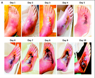
Using graphic pictures taken by a patient who thought a small lesion that was created by the pressure of a new pair of shoes would heal spontaneously, the article documents the infection’s progression.
The obese, 50-year-old patient’s diabetic foot infection began with a swollen, red patch and rapidly evolved into a fully necrotic lesion that ultimately required surgical debridement. According to the article authors, “the preoperative photographs show erythema (day 1), blisters (day 3), a necrotizing abscess (day 6), and wound infection requiring surgery (day 10).”


Following the procedure, the patient was treated with antibiotics for 3 weeks, after which “the infection resolved with no recurrence or sequelae during 3 years of follow-up.”
Based on this photographic case study, the authors concluded, “diabetic foot infection may evolve rapidly, especially in patients with neuropathy.”