
Now Playing
Achondroplasia is driven by overactive FGFR3 signaling that disrupts normal growth plate function. The panel explains how this molecular pathway affects endochondral bone growth and provides the foundation for understanding targeted treatment.
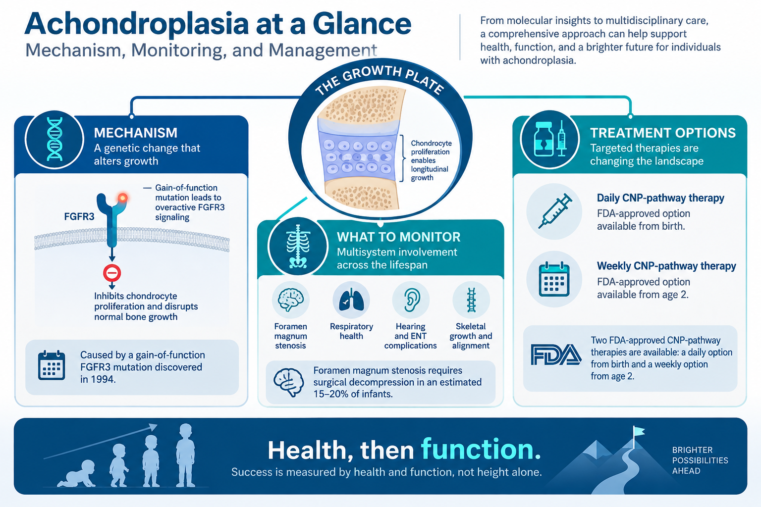
Welcome back to another HCPLive Peer Exchange series. In this episode titled, “Understanding the Pathophysiology of Achondroplasia,” the panel reviews the molecular basis of achondroplasia and the role of fibroblast growth factor receptor 3 (FGFR3) in skeletal growth and development. The discussion begins with the identification of FGFR3 as the gene responsible for achondroplasia and its normal function in regulating chondrocyte proliferation. The panel explains that most appendicular bones and portions of the axial skeleton develop through endochondral ossification, a process that depends on carefully balanced signaling within the growth plate.
Under normal conditions, FGFR3 acts as one of several signals that restrain chondrocyte proliferation. In achondroplasia, a gain-of-function mutation causes FGFR3 signaling to become overactive, creating an excessive inhibitory effect on chondrocyte proliferation and differentiation. This disrupts normal endochondral ossification and leads to impaired longitudinal bone growth and the characteristic skeletal phenotype associated with achondroplasia. By establishing the underlying biology first, the panel provides important context for the remainder of the discussion. Understanding how excessive FGFR3 signaling affects skeletal development also helps explain why contemporary therapies seek to influence this pathway and restore a more favorable balance in growth plate signaling.
In the next episode, “Recognizing and Diagnosing Achondroplasia,” panelists will move from molecular biology to the clinical presentation of the disease. They will discuss the physical and radiographic findings that raise suspicion for achondroplasia and the role of genetic and prenatal testing in confirming the diagnosis.